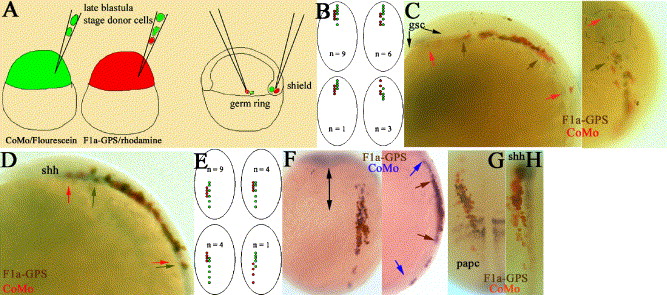
Fig. 6 zfmi1a acts cell-autonomously in axis extension but is not required for dorsal convergence. (A) Schematic representation of transplantation experiments. Donor F1a-GPS1 and CoMo (F1a-GPS1 morpholino but with four nucleotide changes) cells were transplanted simultaneously into the deep cells of the shield or into the germ ring of wild-type shield stage hosts. Only those embryos in which CoMo donor cells had extended extensively within the embryo were considered for analysis, n = 37/60. (B and E) Schematics of the four different types of donor cell movement observed. Dorsal view of 1–3 somite embryos. Red dots: the A-P extension of F1a-GPS1/rhodamine-injected cells; green dots: CoMo/Fluorescein-injected cells. (B) Results of transplants into shield, (E) results of transplants into germ ring. (C, D, F–G) Donor cell transplants into deep cells of shield (C, D) or germ ring (F–G). Representative embryos following whole-mount in situ hybridization and immunostaining. Gene markers are as indicated on the plates. Color of donor cell staining is as indicated on the plates and arrows indicate the limit of extension of each donor population type using the same color coding. (C) Left panel, 3-somite embryo; lateral view of head, anterior is to the left. During immunostaining procedure, posterior prechordal gsc expression has been lost. Right panel, dorsal view showing rostral edge of same embryo, anterior is to top. gsc expression domain within polster is indicated by dashed lines. Arrows indicate rostral boundaries of cell movement (D) 1-somite embryo, lateral view of head. (F) 1-somite embryo, left panel; dorsal view, arrow indicates position of midline. Right panel, lateral view (G and H) Dorsal views of 3-somite embryos.
Reprinted from Developmental Biology, 282(2), Formstone, C.J., and Mason, I., Combinatorial activity of Flamingo proteins directs convergence and extension within the early zebrafish embryo via the planar cell polarity pathway, 320-335, Copyright (2005) with permission from Elsevier. Full text @ Dev. Biol.

