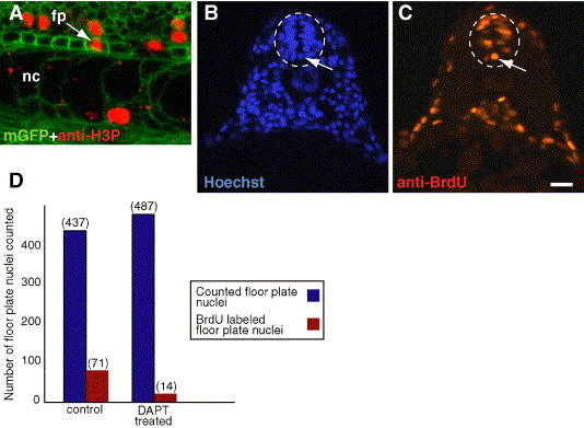
Fig. 6 Notch signaling is required for floor plate proliferation. (A) Side view confocal image, anterior to the left and dorsal up, showing proliferating cells marked by anti-H3P antibody (red) in a 24 hpf Tg(β-actin:mgfp)VU119 embryo. Cells are outlined by expression of a membrane-tethered EGFP. The arrow points to a dividing floor plate cell. (B, C) Both images are from the same transverse section of a 19 hpf (20 somite stage) embryo treated with BrdU to label proliferating cells. (B) Hoechst staining labels all nuclei. Floor plate nuclei (arrow) are easily identifiable and are medially located in the most ventral aspect of the neural tube (white dashed oval). (C) Nucleus labeled by anti-BrdU antibody (arrow) showing that the floor plate cell in panels B was proliferating. (D) We analyzed transverse sections of control embryos treated with BrdU at 19 hpf and embryos treated with DAPT beginning at 16.5 hpf (15-somite stage) and subsequently treated with BrdU at 19 hpf. Blue bars indicate total floor plate nuclei counted in each sample. Red bars indicate the number of floor plate nuclei that were also labeled by anti-BrdU antibody. Scale bar: 13 μm for A; 20 μm for panels B, C.
Reprinted from Developmental Biology, 298(2), Latimer, A.J., and Appel, B., Notch signaling regulates midline cell specification and proliferation in zebrafish, 392-402, Copyright (2006) with permission from Elsevier. Full text @ Dev. Biol.

