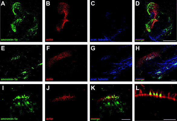
Fig. 2 Expression of anosmin-1a in sensory hair cells of the cristae and in the ampullary nerve of adult zebrafish inner ear. Whole-mount double immunofluorescence with anti-anosmin-1a polyclonal (green) and anti-acetylated tubulin monoclonal (blue) antibodies combined to rhodamin-phalloidin staining of actin (red). (A?D) In the whole cristae, anosmin-1a staining was observed at both extremities of the sensory patch and in the ampullary nerve. (E?H) At the extremity of the cristae, anosmin-1a is accumulated in the ciliated cells, mainly in the hair bundle but also in the cell body, and in the nerve fibers. (I?K) Higher magnification revealed anosmin-1a accumulation in the actin-rich hair bundle and also in the cell body. (L) Confocal section showing anosmin-1a staining of the cilia, in the supranuclear region and at the cell membrane. Data are maximum intensity projections of the acquired z-stacks (A?K) or a single confocal section (L). Scale bar: 50 μm (A?D), 10 μm (E?L).
Reprinted from Gene expression patterns : GEP, 7(3), Ernest, S., Guadagnini, S., Prevost, M.C., and Soussi-Yanicostas, N., Localization of anosmin-1a and anosmin-1b in the inner ear and neuromasts of zebrafish, 274-281, Copyright (2007) with permission from Elsevier. Full text @ Gene Expr. Patterns

