
|
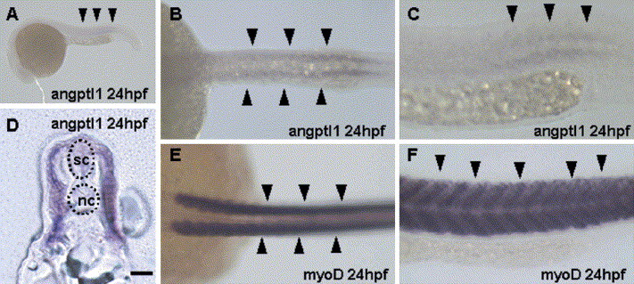
Fig. 2 Embryonic expression of Zangptl1. Whole-mount in situ hybridization was done with a Zangptl (A?D) probe or a myoD probe (E?F) at 24hpf. Weak expression in the somites of a 24hpf embryo especially at the middle portion of tail (arrows in A) was detected. Higher magnification dorsal?ventral (B) and lateral views (C) of a 24hpf embryo are shown. Arrowheads indicate Zangptl1 expression in the somites. (D) A sagittal section of a 24hpf embryo at the middle portion of tail. As comparison of Zangptl1 expression, dorsal?ventral (E) and lateral views (F) of a 24hpf embryo stained with myoD are shown. Arrowheads, somites; sc, spinal cord; nc, notochord. Bar indicates 50 μm.
Reprinted from Gene expression patterns : GEP, 5(5), Kubota, Y., Oike, Y., Satoh, S., Tabata, Y., Niikura, Y., Morisada, T., Akao, M., Urano, T., Ito, Y., Miyamoto, T., Watanabe, S., and Suda, T., Isolation and expression patterns of genes for three angiopoietin-like proteins, Angptl1, 2 and 6 in zebrafish, 679-685, Copyright (2005) with permission from Elsevier. Full text @ Gene Expr. Patterns