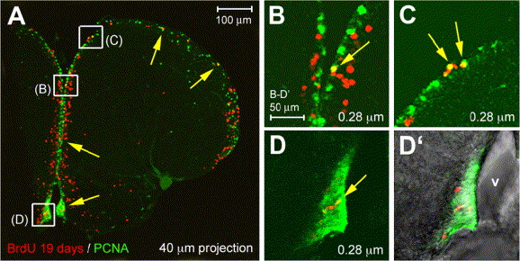
Fig. 7 Long-lasting neuronal progenitors in the zebrafish adult telencephalic ventricular zone. Co-immunodetection of PCNA (green) and BrdU (red) after long-term tracing (19 days), observed under confocal microscopy on cross-sections (thickness indicated, dorsal up). Panels B–D are high magnifications of the areas boxed in panel A, panel D′ is a brightfield and fluorescence view of panel D to locate the ventricle (v). While most labeled cells have exited the ventricular zone, a few BrdU-positive cells are retained in cycle (yellow arrows); these cells are located throughout the DV extent of the telencephalic ventricular zone.
Reprinted from Developmental Biology, 295(1), Adolf, B., Chapouton, P., Lam, C.S., Topp, S., Tannhauser, B., Strähle, U., Gotz, M., and Bally-Cuif, L., Conserved and acquired features of adult neurogenesis in the zebrafish telencephalon, 278-293, Copyright (2006) with permission from Elsevier. Full text @ Dev. Biol.

