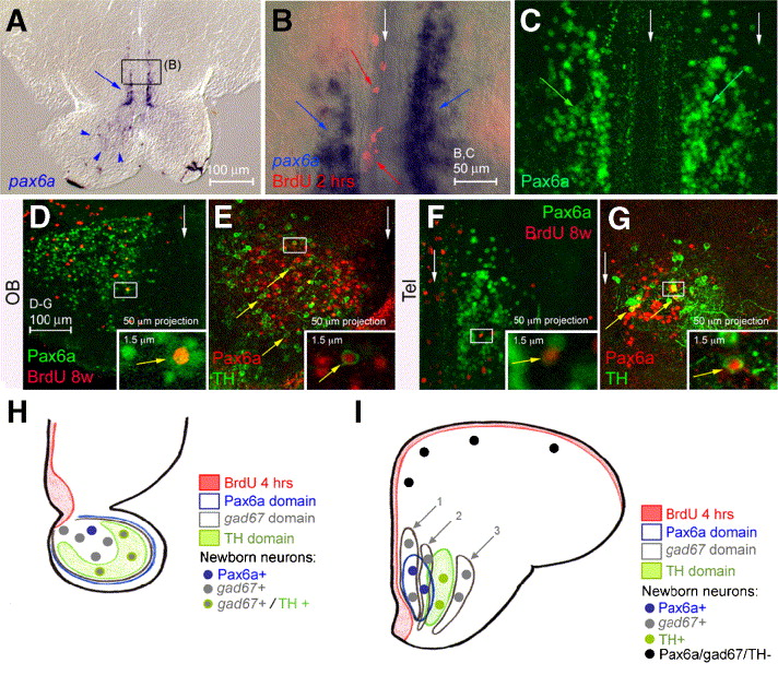
Fig. 6 Pax6a is expressed in neurons of the OB and anterior telencephalon. (A–G) Comparison of pax6a expression (A, B: in situ hybridization, blue; C–G: immunodetection, green or red) with BrdU incorporation (red) after 4 h (B) or 8 weeks (D, F) or with TH (E, G, green). All views are cross-sections, dorsal up, with white arrows to the midline; insets are high magnifications of the boxed areas; panel B is an overlay between bright field and fluorescence. Pax6a is expressed in the OB (A, arrowheads, D,E) and in bilateral telencephalic longitudinal stripes (A, B blue arrows, C green arrows, F, G), but not in proliferating cells (red arrows in panel B, see also C). After 8 weeks of tracing, cells expressing pax6a RNA (not shown) or protein and doubly positive for BrdU can be observed in both the Ob and telencephalon (D, F) (double labeled cells in insets). Coexpression of Pax6a and TH can also be detected in these domains (E, G) (examples of double positive cells are indicated by arrows and some are magnified in insets). (H, I) Schematics summarizing gad67, TH and Pax6a expression in the OB (H) and in the anterior subpallium and pallium (I) as well as the identity of newborn neurons 2 months after BrdU incorporation (color-coded).
Reprinted from Developmental Biology, 295(1), Adolf, B., Chapouton, P., Lam, C.S., Topp, S., Tannhauser, B., Strähle, U., Gotz, M., and Bally-Cuif, L., Conserved and acquired features of adult neurogenesis in the zebrafish telencephalon, 278-293, Copyright (2006) with permission from Elsevier. Full text @ Dev. Biol.

