
|
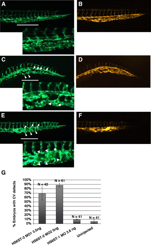
Fig. 4 Zebrafish HS6ST-2 is essential for branching morphogenesis of the caudal vein in zebrafish embryos. (A) Tg(fli-1:EGFP) embryo at 48 hpf under FITC illumination with inset at the lower left corner showing a region of the venous plexus. (B) Angiogram of the Tg(fli-1:EGFP) embryo in (A) under TRITC illumination. (C–D) Tg(fli-1:EGFP) embryo injected with 5 ng of HS6ST-2 MO2 (C) and the angiogram of the same embryo (D) showing reduced branching as indicated by large loops (arrowheads) in the caudal vein plexus. (E–F) In the more severe case, the venous plexus in a Tg(fli-1:EGFP) embryo fails to achieve the complexity as observed in Tg(fli-1:EGFP) embryos. Instead, the formation of a cavernous vessel (arrowheads) is observed. (G) A summary graph showing results of injections using two non-overlapping morpholinos against the zebrafish HS6ST-2 gene. Both oligos generate similar cardinal vein defects including formation of large loops and cavernous vessels with high penetrance. In contrast, HS6ST-1 MO (column 3) does not generate a significant percentage of embryos with CV defects. CV, cardinal vein. N denotes the number of embryos analyzed. ±SEM. The lines in panels A, C and E indicate the segments of venous plexus displayed in insets.
Reprinted from Developmental Biology, 284(2), Chen, E., Stringer, S.E., Rusch, M.A., Selleck, S.B., and Ekker, S.C., A unique role for 6-O sulfation modification in zebrafish vascular development, 364-376, Copyright (2005) with permission from Elsevier. Full text @ Dev. Biol.