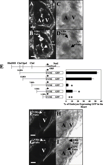
Fig. 5 Distal elements of the GATA-4 promoter are required to direct expression to the atrioventricular valve and caudal aspects of the heart at 6 days post fertilization. (A, B) The 14.8-kb GATA-4 fragment controls GFP expression in the atrioventricular valve (A/V Valve), the bulboventricular valve (V/BA Valve), the ventricle and the atrium. A and B are images at different focal planes to enable visualization of specific regions of the three-dimensional heart. (C, D) Phase of A and B, respectively. (E) GATA-4 promoter analysis. Transient transgenic assays showed that 14.8 or 12 kb of 5′-flanking GATA-4 genomic DNA confers GFP expression to the heart. The HindIII–NotI genomic fragment used to clone the various GATA-4 constructs is schematically represented at the top of the graph. Truncation of 4.2 kb from the 12-kb construct significantly decreased GFP expression in the heart (7.8-kb construct). GFP expression in the heart is lost following further truncation to 1.1 kb of sequence. Statistical analysis was performed between 4 and 6 days post fertilization. Student's t test was performed in comparison to the 12-kb promoter fragment and represents a significant decrease (P < 0.01) in the ability of a specific construct to direct GFP expression in the heart. Significance is indicated by an *. No expression in the heart is indicated by **. Significance could not be determined for constructs that do not express GFP in the heart because the analysis requires standard deviation. However, there is a difference in the expression level between the 1.1-kb construct and the other constructs. ORF = open reading frame. (F, G) Comparable images of A and B except that the transgene contains only 7.8 kb of GATA-4 upstream sequences. Note that expression in the heart is limited to the ventricle and the bulboventricular valve (V/BA valve). F and G are images at different focal planes to enable visualization of specific regions of the three-dimensional heart. (H, I) Phase of F and G, respectively. All images are ventral views with the head at the top of the picture. Scale BAR = 50 μM. A = atrium and V = ventricle.
Reprinted from Developmental Biology, 267(2), Heicklen-Klein, A., and Evans, T., T-box binding sites are required for activity of a cardiac GATA-4 enhancer, 490-504, Copyright (2004) with permission from Elsevier. Full text @ Dev. Biol.

