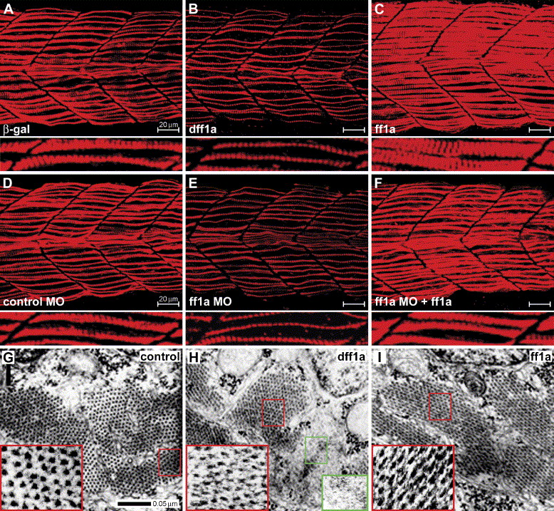
Fig. 5 ff1a functions in slow myofibril assembly. Zebrafish embryos were injected with (A) β-gal (B, H) a deleted ff1a, dff1a (C, I), ff1a (D), control morpholino (MO) (E), ff1a morpholino or (F), ff1a morpholino plus ff1a mRNA, and then their slow myofibrils were stained with F59 antibody. (A?F) Lateral view of slow muscle fibrils in somites 14?17. The figures below them are magnified muscle fibrils. (A) Normal striated fibrils at 24 hpf. (B) Expression of dff1a results in the formation of thinner muscle fibrils. (C) Overexpression of ff1a leads to thicker fibrils with expanded width. (D) Control morpholino-injected embryo does not affect slow fibrils. (E) Embryos injected with ff1aMO have thinner fibrils. (F) Co-injection ff1a mRNA and ff1aMO rescued the phenotype caused by ff1aMO. (G?I) Electron micrographs of the third dorsal slow muscle fiber counting from the pioneer cells of the anal somite. (G) In the control fiber, myosin and actin filaments are organized into sarcomeres with the appearance of arrays of thick and thin black dots inside the sarcomeres (enlarged in the inserted red boxes). (H) In dff1a-injected embryo, myosin thick filaments and actin thin filaments are not well formed (inside red box), a few sarcomeres are not complete (red arrow), and in few sarcomeres, the myofibrils are not formed (green box). (I) Myosin rods are denser in ff1a overexpressed embryos (enlarged in the insert).
Reprinted from Developmental Biology, 286(2), Sheela, S.G., Lee, W.C., Lin, W.W., and Chung, B.C., Zebrafish ftz-f1a (nuclear receptor 5a2) functions in skeletal muscle organization, 377-390, Copyright (2005) with permission from Elsevier. Full text @ Dev. Biol.

