
|
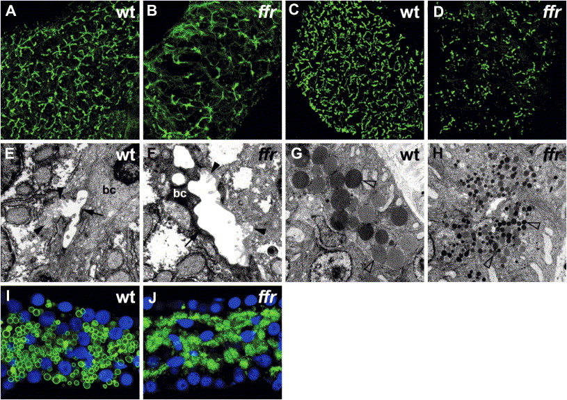
Fig. 1 Phenotypic analysis of ffrmutants A and B) Confocal projections through the liver of 5 dpf wild-type and ffr larvae processed for cytokeratin immunohistochemistry. These immunostainings reveal widespread biliary degeneration in ffr mutants (5 dpf). C and D) Similar immunostainings of 5 dpf wild-type and ffr larvae processed for p-glycoprotein immunohistochemistry. These images of the wild-type and ffr liver show that canalicular architecture is also disrupted in the ffr mutants (5 dpf). E and F) Transmission electron micrographs showed degenerating biliary epithelial cells (bc) (indicated by arrow) and canaliculi (indicated by arrowhead) within the liver of ffr mutants (F) compared with wild-type (E). G and H) Electron micrographs of the pancreas reveal small zymogen granules in the ffr mutants (arrowhead; [H]) compared with wild-type (G). I and J) Confocal image from the pancreas of a 5 dpf ffr mutant (J) processed for carboxypeptidase A immunohistochemistry (green) showed altered distribution of this digestive enzyme compared with wild-type (I). Nuclei stained with Dapi (blue).
Reprinted from Cell Metabolism, 3(4), Ho, S.Y., Lorent, K., Pack, M., and Farber, S.A., Zebrafish fat-free is required for intestinal lipid absorption and Golgi apparatus structure, 289-300, Copyright (2006) with permission from Elsevier. Full text @ Cell Metab.