
|
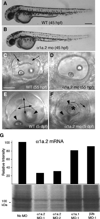
Fig. 3 Knockdown of Na,K-ATPase α1a.2 mRNA disrupts development of semicircular canals. All panels show a lateral view with anterior to the left. Morphants were injected with 2 ng of α1a.2 MO-1. (A) Wild type (WT) embryo at 45 hpf. (B) α1a.2 morphant at 45 hpf. (C) Otic vesicle (OV) of WT embryo at 55 hpf. Arrows indicate protrusions of semicircular canals. (D) OV of α1a.2 morphant at 55 hpf. (E) OV of WT embryo at 5 dpf. Double-headed arrows show hubs of the semicircular canals. Arrowheads indicate otoliths. (F) OV of α1a.2 morphant at 5 dpf. Asterisk shows epithelial mass in center of OV. Arrowheads indicate otoliths. Arrow indicates the posterior crista. (G) Effect of MOs on translation of α1a.2 mRNA. α1a.2 mRNA was translated in the presence of antisense MOs (4 μM) and analyzed as described in Fig. 1A. mo, morphant. Scale bars: A?B, 250 μM; C?F, 50 μM.
Reprinted from Developmental Biology, 294(1), Blasiole, B., Canfield, V.A., Vollrath, M.A., Huss, D., Mohideen, M.A., Dickman, J.D., Cheng, K.C., Fekete, D.M., and Levenson, R., Separate Na,K-ATPase genes are required for otolith formation and semicircular canal development in zebrafish, 148-160, Copyright (2006) with permission from Elsevier. Full text @ Dev. Biol.