Image
Figure Caption
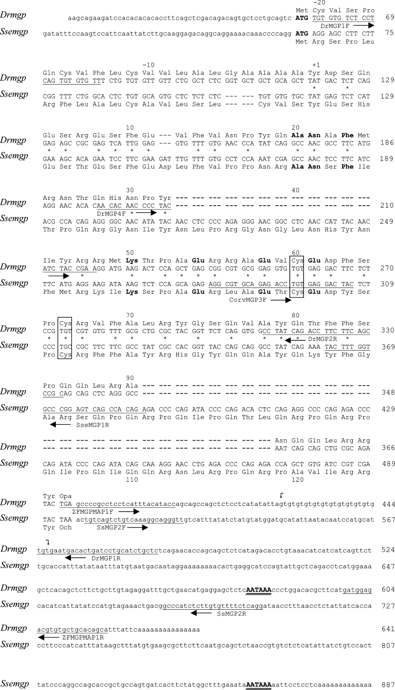
Fig. S2 Zebrafish and sole mgp cDNAs. Nucleotide sequence of the two mgp cDNAs were obtained by a combination of RT- and 5′ RACE-PCR amplification. Numbering and labeling is as described in Figure 2s. Amino acid residues are numbered according to the first residue of the mature protein and are shown above the corresponding codon in the DNA sequence, for zebrafish. A 13 (GT) repetitive motif on zebrafish mgp 3′-UTR region is marked between curved arrows.
Acknowledgments
This image is the copyrighted work of the attributed author or publisher, and
ZFIN has permission only to display this image to its users.
Additional permissions should be obtained from the applicable author or publisher of the image.
Reprinted from Gene expression patterns : GEP, 6(6), Gavaia, P.J., Simes, D.C., Ortiz-Delgado, J.B., Viegas, C.S., Pinto, J.P., Kelsh, R.N., Sarasquete, M.C., and Cancela, M.L., Osteocalcin and matrix Gla protein in zebrafish (Danio rerio) and Senegal sole (Solea senegalensis): Comparative gene and protein expression during larval development through adulthood, 637-652, Copyright (2006) with permission from Elsevier. Full text @ Gene Expr. Patterns

