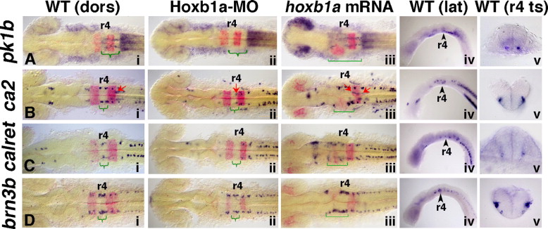
Fig. 4
Fig. 4 Expression of validated Hoxb1a target genes with neuronal specific expression domains within r4. In situ hybridization at 20 hpf demonstrates that pk1b, ca2, calret and brn3b are regulated by Hoxb1a. Anterior to the left in all panels. Rhombomere (r) 4 is labeled. krox20 marks r3 and r5 (panels i?iii; in red). Curved green brackets indicate Hoxb1a-dependent expression (compare panels i with ii). Square green brackets indicate expanded Hoxb1a-dependent expression in panel iii. Panels i and iv: (A) In wild-type embryos, pk1b is expressed in narrow bilateral trails through r5 and r6 (green bracket) as well as at low levels in ventral r4. (B) ca2 is expressed in bilateral discrete domains in r4, r5, and r6 in wild-type embryos (green bracket, Hoxb1a-dependent expression in r4), as well as in the trigeminal (r2) and facial (r5/6; red arrows) BMNs. (C) calret is expressed bilaterally in the hindbrain in three domains in wild-type embryos (green bracket, Hoxb1a-dependent expression in r4). (D) brn3b is expressed bilaterally in r3?r5 in wild-type embryos, with an elevated expression domain in r4 (green bracket, Hoxb1a-dependent expression). Panel ii: The r4-specific expression of all four genes is missing or reduced in Hoxb1a-MO-injected embryos (green brackets). Note that in panel Bii, while the four Hoxb1a-dependent domains are missing from r4, the Hoxb1a-independent ca2 expression is retained in the facial BMNs, which fail to migrate from r4 in Hoxb1a-deficient embryos (red arrow). Panel iii: The r4-specific expression of all four genes is anteriorly expanded in hoxb1a mRNA-injected embryos (square green brackets). Note that in panel Biii, the bilateral Hoxb1a-dependent ca2 expression is expanded anteriorly (square green brackets), and the Hoxb1a-independent expression of ca2 continues to mark the facial BMNs which have also expanded anteriorly (red arrows). Panel v: Transverse sections through wild-type embryos show that (A) pk1b is expressed in discrete bilateral ventral cells in r5 and at low levels throughout ventral r4 (section includes both r4 and r5). (B?D) Hoxb1a-dependent expression of ca2, calret, and brn3b is found laterally within the ventral half of the neural tube. dors: dorsal; lat: lateral; MO: morpholino; ts: transverse.
Reprinted from Developmental Biology, 309(2), Rohrschneider, M.R., Elsen, G.E., and Prince, V.E., Zebrafish Hoxb1a regulates multiple downstream genes including prickle1b, 358-372, Copyright (2007) with permission from Elsevier. Full text @ Dev. Biol.

