
|
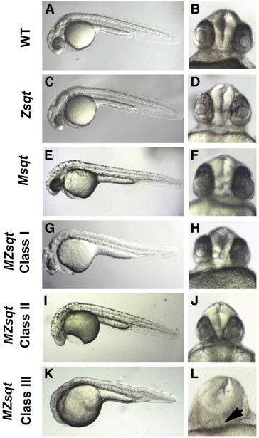
Fig. 1 Embryos lacking maternal sqt function have variable defects. Lateral (A, C?E, G, I, K) or ventral (B, D, F, H, J, L) images of live embryos at 30 hpf. The body axis forms normally in wild type (A), Zsqt (C) and Msqt (E) embryos, and none of these embryos are cyclopic (B, D, F), although Zsqt mutants from some crosses display mild cyclopia. Embryos lacking both maternal and zygotic sqt can be classified into three groups depending on their phenotype. The frequency of the phenotypes depends on the genetic background of the mother, as described in the text. The MZsqt embryos depicted in panels G?L are siblings. Class I MZsqt mutants (G, H) are indistinguishable from wild type, while Class II mutants (I, J) display mild cyclopia (J), typical of the previously reported phenotype of Zsqt mutants (Dougan et al., 2003). Class III mutants have reduced dorsal and anterior structures, characterized by reduced (L, arrow) or absent eyes. Anterior is to the left when apparent.
Reprinted from Developmental Biology, 309(2), Hagos, E.G., Fan, X., and Dougan, S.T., The role of maternal Activin-like signals in zebrafish embryos, 245-258, Copyright (2007) with permission from Elsevier. Full text @ Dev. Biol.