
|
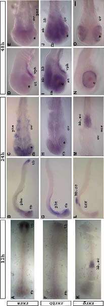
Fig. 4 Expression of rxra (A-E), rxrbb (F-J) and rxrg (K-O) at 12 hpf (A,F,K), 24 hpf (B,C,G,H,L,M) and 48 hpf (D,E,I,J,N,O). Refer to text for detailed descriptions. Flat-mount embryos seen from a dorsal view (A,C,E,F,H,J,K,M,O) or a lateral view (B,D,G,I,L,N). Anterior is to the left. Abbreviations: e, eye; fb, forebrain; hb, hindbrain; mb, midbrain; ncr, neural crest; phe, pharyngeal endoderm; ov, otic vesicle; sc, spinal cord; st, stomadeum; tb, tailbud; vph, ventral tissue in pharyngeal region. Scale bar represents 115 μm in 4A,F,K; 100 μm in 4B,G; 108 μm in 4L; 40 μm in 4C,M; 35 μm in 4H; 32 μm in 5D,E,I,J,N,O.
Reprinted from Gene expression patterns : GEP, 6(5), Tallafuss, A., Hale, L.A., Yan, Y.L., Dudley, L., Eisen, J.S., and Postlethwait, J.H., Characterization of retinoid-X receptor genes rxra, rxrba, rxrbb and rxrg during zebrafish development, 556-565, Copyright (2006) with permission from Elsevier. Full text @ Gene Expr. Patterns