
Image
|
Figure Caption
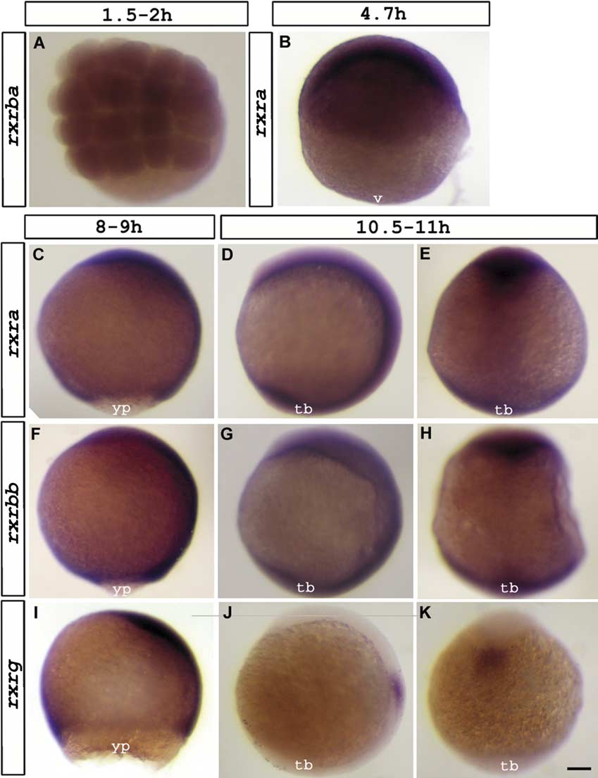
Fig. 3 Expression of rxra (B, C-E), rxrba, (A), rxrbb (F-H) and rxrg (I-K) visualized at 1.5-2 hpf (A), 4.7 hpf (B), 8-9 hpf (C,F,I) and 10.5-11 hpf (D,E,G,H,J,K). Dorsal view (A,E,H,K). Lateral view; anterior up and dorsal to the left (B,C,D,F,G,I,J). Abbreviations: v, vegetal pole; yp, yolk plug; tb, tailbud. Scale bar represents 93 μm in A; 106 μm in B, C-K.
Figure Data
Acknowledgments
This image is the copyrighted work of the attributed author or publisher, and
ZFIN has permission only to display this image to its users.
Additional permissions should be obtained from the applicable author or publisher of the image.
Reprinted from Gene expression patterns : GEP, 6(5), Tallafuss, A., Hale, L.A., Yan, Y.L., Dudley, L., Eisen, J.S., and Postlethwait, J.H., Characterization of retinoid-X receptor genes rxra, rxrba, rxrbb and rxrg during zebrafish development, 556-565, Copyright (2006) with permission from Elsevier. Full text @ Gene Expr. Patterns