Image
Figure Caption
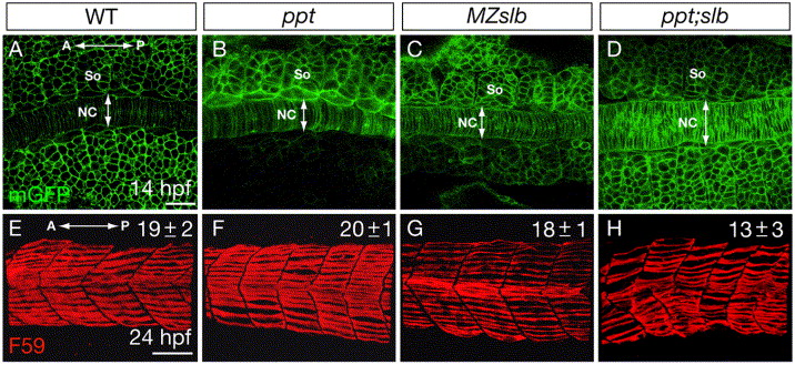
Fig. S2 Fig. S2. The somite morphology and slow muscle development in the non-canonical Wnt/PCP mutants. (A-D) Confocal images of the embryos expressing mGFP at the 10-somite stage, dorsal views. (E-H) Lateral views of day1-embryos stained with F59 antibody that labels the slow muscle fibers. The numbers of slow muscle fibers per somite are indicated in the up right corner. All the measurements were done in the 3rd and 4th myotomes.10 embryos were analyzed for each genotype. NC, notochord. So, somite. A, anterior. P, posterior. Scale bar, 50 μm.
Figure Data
Acknowledgments
This image is the copyrighted work of the attributed author or publisher, and
ZFIN has permission only to display this image to its users.
Additional permissions should be obtained from the applicable author or publisher of the image.
Reprinted from Developmental Biology, 304(1), Yin, C., and Solnica-Krezel, L., Convergence and extension movements mediate the specification and fate maintenance of zebrafish slow muscle precursors, 141-155, Copyright (2007) with permission from Elsevier. Full text @ Dev. Biol.

