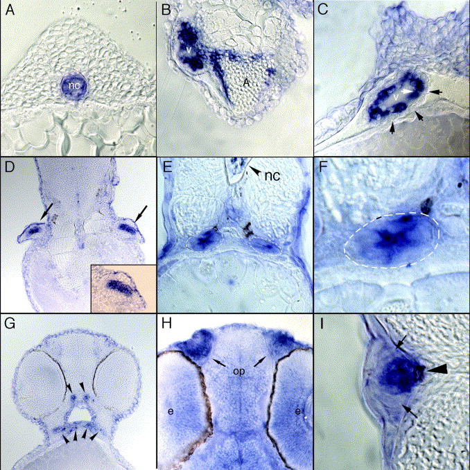
Fig. 3 Histological analysis of trpv4 expression. Embryos used for in situ hybridization analysis were embedded in JB-4 and sectioned at 10–12.5 μm then examined by light microscopy. (A) Expression is restricted to the notochord in 18 hpf embryos. (B) Sections through a 72 hpf embryo heart show strong expression in the ventricle (v) and upper regions of the atrium (a). (C) A closer examination of ventricular staining illustrates that trpv4 mRNA expression is restricted to cells of the endocardium (white arrow) and absent from surrounding myocardial cells (black arrows). (D) Frontal section of 52 hpf embryos highlighting the presence of trpv4 mRNA in the forming cartilage of the pectoral fin buds (black arrows and magnification in inset). (E, and F) trpv4 expression in the pronephric duct (white dashes) is detected in 32 hpf embryos. (G) Cross section through 52 hpf embryos shows expression in the developing bones of the jaw (arrowheads). (H) Frontal section through a 32 hpf embryo shows that trpv4 mRNA is strongly expressed throughout the brain and eye (e) with enhanced expression in the olfactory placodes (op). (I) Cross section through a lateral line organ of a 32 hpf embryo shows specific expression in hair cell bodies of the neuromast (arrowhead) and no expression in surrounding mantle cells (small arrows).
Reprinted from Gene expression patterns : GEP, 7(4), Mangos, S., Liu, Y., and Drummond, I.A., Dynamic expression of the osmosensory channel trpv4 in multiple developing organs in zebrafish, 480-484, Copyright (2007) with permission from Elsevier. Full text @ Gene Expr. Patterns

