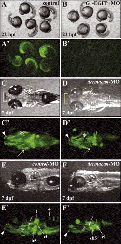
Fig. 7 Effects of dermacan morpholino on external morphology and bones at 7 dpf. (A–B′) Downregulation of dermacan G1–EGFP fusion protein translation by dermacan-MO. (A,A′) Control embryos at 22 hpf injected with 20 pg of dermacan G1–EGFP mRNA in the bright field (A) and dark field (A′). Notice the EGFP signals in all embryos (A′). (B, B′) Embryos at 22 hpf co-injected with 20 pg of dermacan G1–EGFP mRNA and 8 ng of dermacan-MO in the bright field (B) and dark field (B2). Note that EGFP signals were completely suppressed in all embryos. (C–F′) dermacan gene targeting by antisense morpholino. (C,C′,E,E′) Embryos injected with control-MO. (D,D′,F,F′) Embryos injected with dermacan-MO. (C′,D′,E′,F′) Bones were fluorescently labeled with Calcein (visualized by green color). (C,C′,E,E′) control-MO injected larvae developed normally. (E′) Vertebrae numbers 1 and 4 were calcified. (D,D′,F,F′) dermacan-MO injected larvae showed several abnormalities. (D,F) Jaw (basket) and gill cover (dotted line) were morphologically defective. (D′,F′) Note that the dentary (arrowheads), opercle (arrows), and branchiostegal ray (asterisks) were either malformed or absent. Vertebrae were not detected in F′. Anterior to the left. (C,C′,D,D′) Ventral views. (E,E′,F,F′) Lateral views. Abbreviations: cb5, ceratobranchial 5; cl, cleithrum; e, eye; ov, otic vesicle; y, yolk.
Reprinted from Mechanisms of Development, 121(3), Kang, J.S., Oohashi, T., Kawakami, Y., Bekku, Y., Izpisua, B.J.C., and Ninomiya, Y., Characterization of dermacan, a novel zebrafish lectican gene, expressed in dermal bones, 301-312, Copyright (2004) with permission from Elsevier. Full text @ Mech. Dev.

