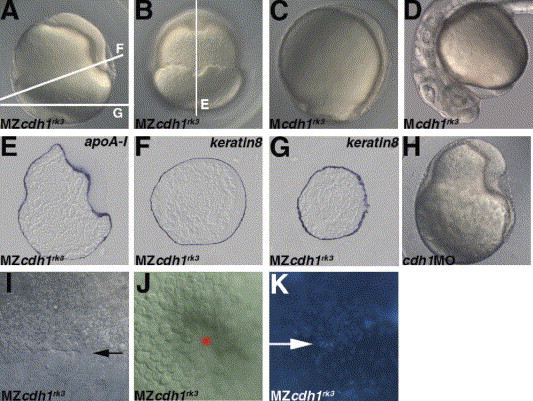
Fig. 4 Maternal-zygotic (MZ) cdh1rk3 mutant embryos show epiboly arrest. (A, B) MZcdh1rk3 mutant embryos displayed epiboly arrest at 10 hpf. MZcdh1rk3 mutant embryos were obtained by crossing the homozygous cdh1rk3 fish, which were rescued by mcdh1 RNA injection. Lateral views with dorsal to the right (A). Dorsal views (B). (C, D) Maternal (M) cdh1rk3 mutant embryos displayed a mild epiboly delay at 10 hpf (C) and normal anterior neural tissues and hatching glands (D). Mcdh1rk3 mutant embryos were obtained by crossing the homozygous cdh1rk3 mutant fish and wild-type fish. Lateral views with dorsal to the right (C). Lateral views in the anterior region (D). (E?G) Expression of the yolk syncytial marker apoA-I (E) and enveloping layer marker keratin8 (F, G) at 10 hpf in the MZcdh1rk3 mutant embryos. The stained MZcdh1rk3 mutant embryos were sectioned as indicated by the white lines in A and B. (H) Morphology of the embryos that received 50 pg of cdh1MO (recognizing the splicing donor of the first intron) at 10 hpf. The cdh1 morphant embryos showed a phenotype similar to that of MZcdh1rk3 mutant embryos. (I, J) DIC images of the margin of the deep cells (I) (marked by an arrow) and enveloping layer (J) at the vegetal pole (marked by a star) in the MZcdh1rk3 mutant embryos at 10 hpf. Lateral views (I) and vegetal pole views (J). The enveloping layer but not the deep cells reached the vegetal pole. (K) DAPI staining of 10 hpf MZcdh1rk3 mutant embryos. Dorsal views. The margin of the deep cells is indicated by an arrow. On the animal pole side, there was a high density of DAPI-stained nuclei, indicating the deep cells. On the vegetal pole side, the nuclei were distributed sparsely, indicating the yolk syncytial layer.
Reprinted from Mechanisms of Development, 122(6), Shimizu, T., Yabe, T., Muraoka, O., Yonemura, S., Aramaki, S., Hatta, K., Bae, Y.K., Nojima, H., and Hibi, M., E-cadherin is required for gastrulation cell movements in zebrafish, 747-763, Copyright (2005) with permission from Elsevier. Full text @ Mech. Dev.

