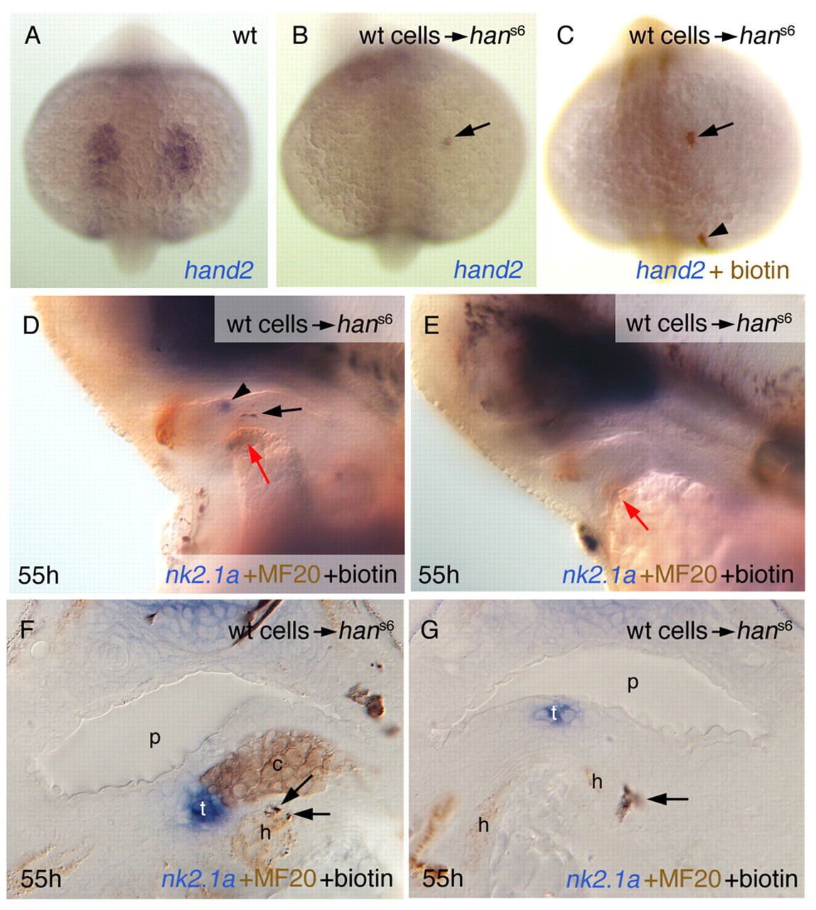
Fig. 4 Grafted wild-type cells can restore nk2.1a expression in hans6 mutant embryos. Labelling of panels is as in Fig. 1. Anterior up (A-C) or to the left (D,E), and sections (F,G). (A-C) Grafted wild-type cells express han (hand2) in the mutant background. Arrows point to a single grafted cell that contributed to the anterior lateral plate mesoderm. The arrowhead in C indicates a grafted cell that contributed to other tissue, not expressing hand2. (D-E) Examples of hosts fixed at 55 hours post fertilisation (hpf). D shows an embryo (corresponding to #4 in Table 1) that has restored nk2.1a expression (arrowhead), an occurrence that was never seen in siblings. The black arrow points to grafted wild-type cells, the red arrows to the heart rudiment. E shows a hans6 embryo without restored thyroidal nk2.1a expression. (F,G) Cross sections of specimens of hans6 embryos with restored nk2.1a expression. Grafted cells in the embryo shown in F (corresponding to #5 in Table 1) are adjacent to the thyroid in cartilage, but also in heart tissue (arrows). Note also that such a large thyroid as in F was never observed in untreated hans6 embryos. Often, donor cells clearly belong to the pharyngeal mesenchyme, but are also close to the heart rudiment (G, embryo corresponds to #3 in Table 1). c, cartilage precursor cells; h, heart rudiment; p, pharynx; t, thyroid.
Image
Figure Caption
Acknowledgments
This image is the copyrighted work of the attributed author or publisher, and
ZFIN has permission only to display this image to its users.
Additional permissions should be obtained from the applicable author or publisher of the image.
Full text @ Development

