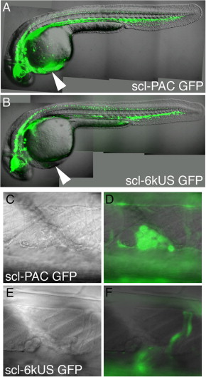
Fig. 7 The second GFP transgenic line, scl-6kUS-GFP lacks elements necessary for expression in definitive hematopoietic clusters. (A) 34 hpf scl-PAC-GFP embryo, and (B) 34 hpf scl-6kUS-GFP embryo, lateral views. In both embryos, GFP is expressed in the CNS and endothelium in identical patterns. In panel A, expression in the primitive erythrocytes is easily seen as they pass over the yolk cell and enter the heart (arrowhead). In panel B, there is much reduced expression in erythroid cells, but is still faintly visible (see also, panel F). (C and D) 4 dfp scl-PAC-GFP embryo, mid-trunk hematopoietic cluster. A typical cluster of approximately 15 cells is visible in the DIC image (C). Strong expression of GFP in these cells is clearly visible (overlay of fluorescent and DIC image) (E and F), 4 dfp scl-6kUS-GFP embryo, mid-trunk hematopoietic cluster. A cluster of approximately 13 cells is visible in the DIC image (E). No expression of GFP is visible in the cluster (overlay of fluorescent and DIC image). Faint expression in circulating primitive erythrocytes is visible in the lumen of the vessels.
Reprinted from Developmental Biology, 307(2), Zhang, X.Y., and Rodaway, A.R., SCL-GFP transgenic zebrafish: In vivo imaging of blood and endothelial development and identification of the initial site of definitive hematopoiesis, 179-194, Copyright (2007) with permission from Elsevier. Full text @ Dev. Biol.

