
|
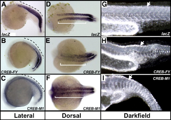
Fig. 7 Distribution of myoD RNA in the trunk and tail of injected embryos is disrupted in embryos with perturbed CREB function. Embryos injected with lacZ (A, D), CREB-FY (B, E) or CREB-M1 (C, F) were stained by WISH during late somitogenesis (22–24 somite stage). CREB-FY injected embryos exhibit less MyoD staining in the trunk somites, 1–15, (dashed line, B; bar, E) than do CREB-M1 (C, F) or control (A, D) embryos. MyoD staining in the posterior-most 7–9 (tail) somites is relatively unchanged. Darkfield microscopy shows somite morphogenesis (arrows, G–I) is varyingly disrupted in both CREB-FY (H) and CREB M1 (I) injected embryos relative to controls (G), which display regularly-spaced, chevron-shaped somites. CREB-FY injected fish display irregularly spaced and sized somites, although the basic chevron-shape is retained (H). CREB-M1 injected fish show additional loss of somite structure and shape (I; note U-shaped somites).
Reprinted from Developmental Biology, 307(1), Dworkin, S., Heath, J.K., Dejong-Curtain, T.A., Hogan, B.M., Lieschke, G.J., Malaterre, J., Ramsay, R.G., and Mantamadiotis, T., CREB activity modulates neural cell proliferation, midbrain-hindbrain organization and patterning in zebrafish, 127-141, Copyright (2007) with permission from Elsevier. Full text @ Dev. Biol.