
|
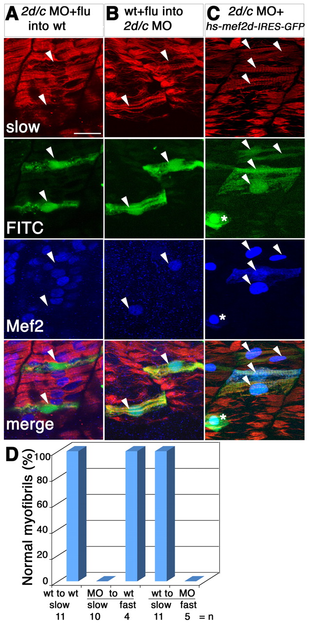
Fig. 4 Mef2 controls slow fibre myofibril assembly cell-autonomously and rescues the morphant. Detection of slow MyHC (red), Mef2 (blue) and FITC or GFP (green) in manipulated 24-hpf zebrafish embryos. (A,B) Transplantation of FITC-dextran-labelled cells (arrowheads, green) from mef2d/c morphants into wild-type host (A) or from wild-type donor into mef2d/c morphant host (B). Note the immature character of the FITC-labelled transplanted fibres in A and the greater maturity of transplanted cells in B, compared with their neighbours. (C) Co-injection of hs-mef2d-IRES-GFP and mef2d/c MO followed by heat shock at 22 hpf rescues myofibril structure in the GFP-expressing cells, which also contain nuclear Mef2 (arrowheads). Asterisk indicates a Mef2-positive skin cell. (D) Bar chart showing quantification of transplantation results.