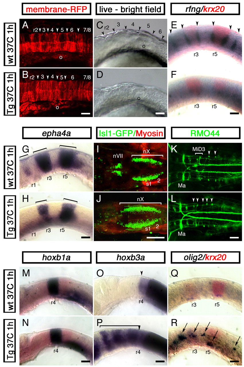
Fig. 6 Cdx4 overexpression in the zebrafish hindbrain disrupts rhombomere formation and promotes spinal cord development. Wild-type embryos (A,C,E,G,I,K,M,O,Q) and their siblings carrying one copy of a phsp70:cdx4 transgene (B,D,F,H,J,L,N,P,R) were heat shocked for 1 hour at 37°C at the three-somite stage and then grown at 28°C for a total of 16 (14 somites, A,B), 19 (20 somites, C-H,M-R) or 50 (I-L) hours. (A,B) Confocal images of the hindbrain region of 14-somite stage embryos injected with membrane-anchored RFP (gap43-RFP) mRNA to reveal rhombomere furrow formation (arrowhead). In all 12 embryos examined, cdx4 overexpression impaired furrow formation. (C,D) Distinctive rhombomere bulges (arrowheads in C) fail to appear in cdx4-overexpressing embryos (D) at the 20-somite stage (n=15). (E,F) Embryos overexpressing cdx4 fail to express the rhombomere boundary marker rfng (purple, arrowheads). krx20 expression was used to visualize r3 and r5 (red). (G,H) Loss of epha4a expression in r1 and reduction in r3 and r5 (brackets) in embryos overexpressing cdx4. (I,J) cdx4 overexpression results in the rostral expansion of vagal (nX) motor neurons and the loss of facial (nVII) and trigeminal (nV, not shown) motor neurons in isl1-GFP embryos. (K,L) In wild-type embryos, the RMO44-positive MiD3 reticulospinal neurons are found forming a cluster in r6 (brackets, K). This cluster is replaced by individual r7/8-like, T reticular interneurons in cdx4-overexpressing embryos (arrowheads, L). On a few occasions, MiD2 (r5) and Mauthner (r2) neurons (Ma) were also lost in cdx4-overexpressing embryos (data not shown). (M,N) Expression of the r4 marker hoxb1a is not affected in embryos overexpressing cdx4. (O,P) hoxb3a, a gene that is normally transcribed in rhombomeres posterior to r4 (O, arrowhead), is expressed throughout the rostral hindbrain in cdx4-overexpressing embryos (bracket in P). (Q,R) cdx4 overexpression induces ectopic hindbrain expression of the spinal motor neuron and oligodendrocyte marker olig2 (purple, arrows in R). For the 20-somite stage embryos (C-H,M-R), a minimum of 36 embryos from at least three independent experiments were analyzed. All embryos mounted anterior to the left, lateral views except for A,B,I-L, which are dorsal. o, otic vesicle; r, rhombomere; s, somite. Scale bars: 100 µm.
Image
Figure Caption
Acknowledgments
This image is the copyrighted work of the attributed author or publisher, and
ZFIN has permission only to display this image to its users.
Additional permissions should be obtained from the applicable author or publisher of the image.
Full text @ Development

