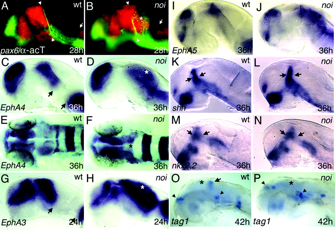
Fig. 3 Marker analysis in wild-type (wt) and noi mutant embryos suggests a respecification of midbrain tissue. In situ hybridisation with the indicated marker genes at the given stages (h, hours postfertilisation), lateral views (except dorsal views in E, F) with anterior to the left; asterisks mark the position of the misspecified midbrain. A: Staining for pax6.1, a marker for the forebrain and basal hindbrain, combined with an acetylated tubulin staining to visualize outgrowing axons. The posterior commissure (PC, marked by white arrowheads) is located at the diencephalic-mesencephalic boundary in the pax6.1+ domain. B: In the noi mutant, pax6.1 expression is observed in the territory of the presumptive midbrain and new branches of the PC (yellow arrowheads) are visible more posterior to the endogenous position. Similar to the forebrain, the hindbrain expression domain expands anteriorly into the midbrain territory (A,B, white arrows). Markers of the Ephrin family show a similar phenomenon: EphA4 (C,E), EphA3 (G), and EphA5 (I) respect the boundary between the diencephalon and the mesencephalon (black arrows). D,F,H,J: In the noi mutant, the expression expands into the misspecified midbrain area. K-N: Marker genes of the zona limitans intrathalamica, such as shh and nkx2.2, show no difference between noi mutant and wild-type siblings, suggesting a correct formation of this anterior region between the prosomeres 2 and 3 (arrows). O: tag1 marks a subset of developing neurons in the cerebellar anlage (arrow). P: This expression domain is missing in the noi mutants, consistent with the loss of cerebellar identity in the noi mutants. However, the expression domains in the dorsal forebrain and in the anterior hindbrain are not altered (indicated by arrowheads).
Image
Figure Caption
Acknowledgments
This image is the copyrighted work of the attributed author or publisher, and
ZFIN has permission only to display this image to its users.
Additional permissions should be obtained from the applicable author or publisher of the image.
Full text @ Dev. Dyn.

