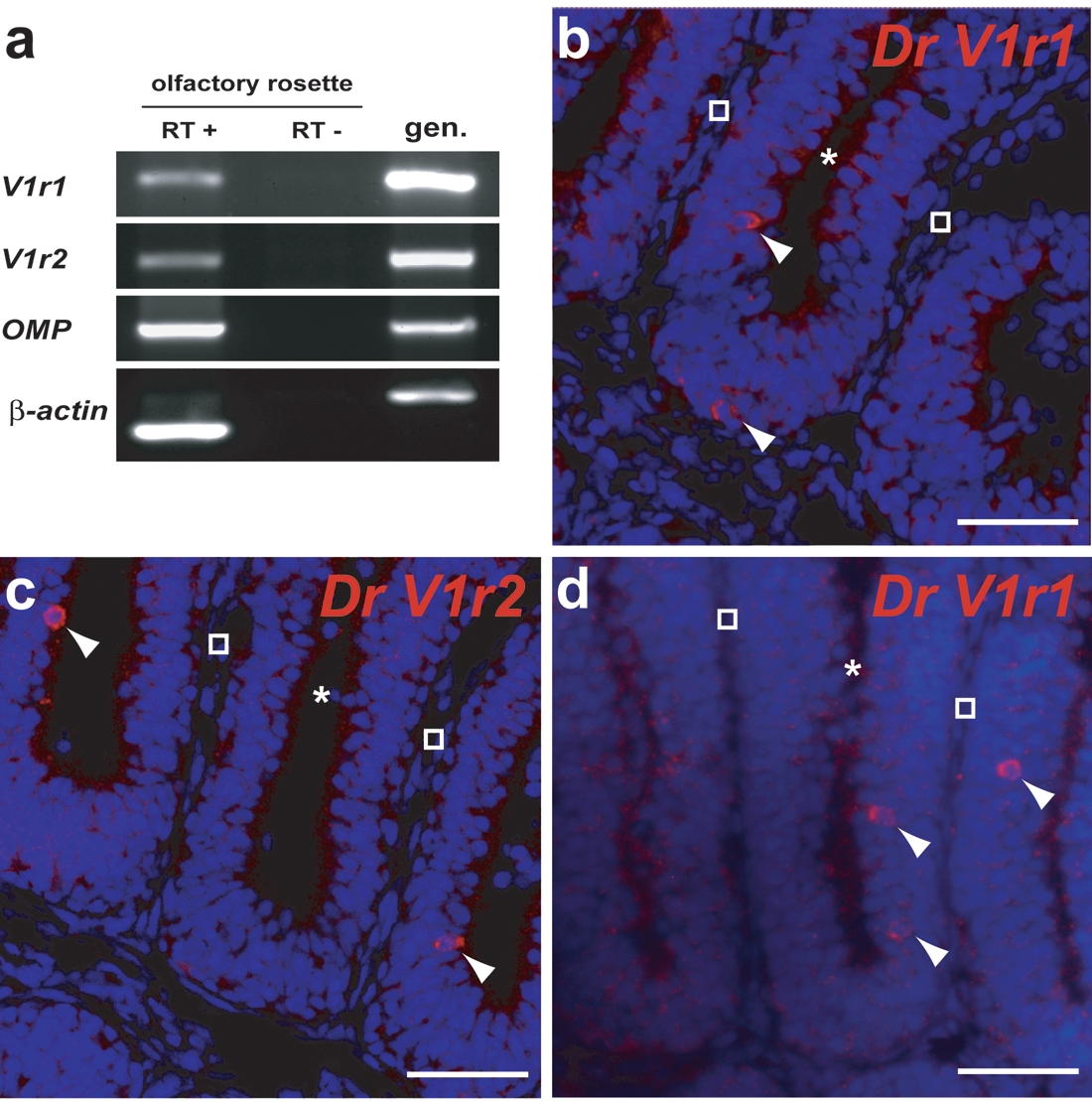
Fig. 5 Dr V1r1 and V1r2 are expressed in the olfactory rosette. (a) RT-PCR indicating transcription of V1r1 and V1r2 in olfactory rosette extracts. OMP (olfactory marker protein) and βactin were used as positive controls. (b) In situ hybridization of a horizontal olfactory rosette section with an anti-sense Dr V1r1 probe (probe III in Figure 3). (c) In situ hybridization with anti-sense Dr V1r2 probes (probe I in Figure 3). (d) In situ hybridization with an antisense 52UTR V1r1 probe (probe II in Figure 3). Arrows indicate cells reacting to the probes. Asterisks and empty squares correspond respectively to luminal and cartilaginous parts of the rosette. Scalebar: 40 μm.
Image
Figure Caption
Figure Data
Acknowledgments
This image is the copyrighted work of the attributed author or publisher, and
ZFIN has permission only to display this image to its users.
Additional permissions should be obtained from the applicable author or publisher of the image.
Full text @ PLoS One

