
|
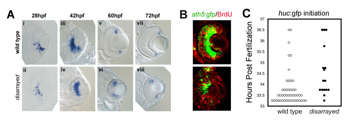
Fig. 4 Initiation of retinal neurogenesis in disarrayed eyes. (A) In situ analysis of ath5 expression during retinogenesis in wild-type (i, iii, v, vii) and disarrayed eyes (ii, iv, vi, viii). (B) BrdU pulse-labeling (red) in wild-type (upper) or disarrayed (lower) embryos carrying the ath5:gfp transgene (green). Embryos were injected with BrdU at 36 hpf and fixed at 42 hpf. Note that in both genotypes, ath5:GFP expression in central retinal cells is post-S-phase. (C) Developmental time (hours post fertilization) for the initiation of huc:GFP expression. The initiation of GFP expression was recorded for all embryos at 0.5 hr intervals in a clutch from an in-cross of disarrayed heterozygous parents (one circle represents one embryo; mutant (black circles) and wild-type (white circles)). Note that all embryos initiate huc:GFP expression within the same window of development. Results from one representative experiment (n = 16 mutant and n = 45 wild-type embryos from one clutch of embryos).