
|
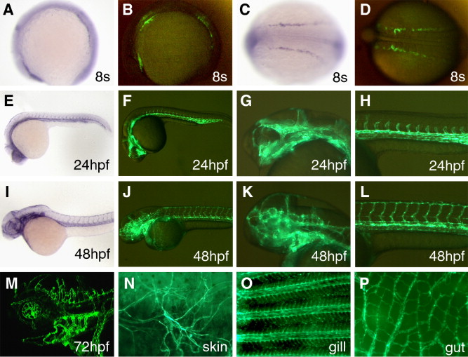
Fig. 1 GFP expression in TG(flk1:GFP)la116 embryos. (A?D) GFP signals driven by the flk(6.4)-GFP transgene can be detected in TG(flk1:GFP)la116 embryos as early as the 8-somite stage. flk1 expression is detected in three patches of cells by in situ hybridization (A). A similar pattern is seen in TG(flk1:GFP)la116 embryos (B). Lateral views are shown in panels A and B and the dorsal views are shown in panels C and D. Anterior to the left. (E?H) GFP expression pattern of TG(flk1:GFP)la116 embryos at 24 hpf (F) resembles the flk1 pattern detected by in situ hybridization (E). Higher magnification images of the head and trunk are shown in panels G and H, respectively. (I?L) GFP expression pattern of TG(flk1:GFP)la116 embryos at 48 hpf (J) resembles the flk1 pattern detected by in situ hybridization (I). Higher magnification images of the head and trunk are shown in panels K and L, respectively. (M) Confocal image of GFP expression in endothelial cells in the brain and brachial arches in 3-day-old TG(flk1:GFP)la116 embryos. (N?P) GFP expression remains active in adult TG(flk1:GFP)la116 fish. Images show GFP signals in endothelial cells in the skin (N), gill (O) and gut (P).
Reprinted from Developmental Biology, 304(2), Choi, J., Dong, L., Ahn, J., Dao, D., Hammerschmidt, M., and Chen, J.N., FoxH1 negatively modulates flk1 gene expression and vascular formation in zebrafish, 735-744, Copyright (2007) with permission from Elsevier. Full text @ Dev. Biol.