
|
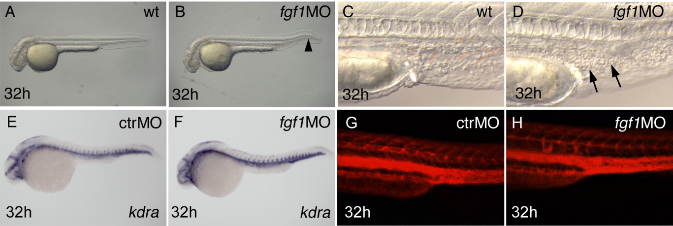
Fig. 2 fgf1 morphants show accumulation of blood cells in the posterior intermediate cell mass (ICM), despite apparently normal vessel pattern and initially normal hematopoiesis. Stages are indicated bottom left, manipulation of embryos top right, and marker expression bottom right. A-D,G,H: Live embryos. Occasionally, fgf1 morphants have a kinked tail (arrowhead in B). Arrows in D point to accumulating cells in the ICM. E,F: The vessel marker kdra is normally expressed. G,H: Microangiography results in a grossly normal pattern of fgf1 morphants (rhodamine-dextran fluorescence of injected embryos, trunk, lateral view).