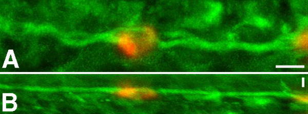
Fig. 2 Close association of neurons expressing spt with the dorsal longitudinal fasciculus. Images shown are projections of serial 0.5 μm optical sections through a 22 hpf embryo stained to reveal spt transcripts (red) and acetylated tubulin (green) that marks axons. The cell shown lies in that part of the developing spinal cord midway along the yolk extension. Rostral is to the left in both images. A shows a lateral projection with dorsal to the top. B shows a dorsal projection with medial to the bottom and lateral to the top. The size bar in A indicates 10 μm. B has an identical rostrocaudal dimension but the mediolateral dimension is compressed. The size bar in B indicates 10 μm in the mediolateral dimension.
Image
Figure Caption
Figure Data
Acknowledgments
This image is the copyrighted work of the attributed author or publisher, and
ZFIN has permission only to display this image to its users.
Additional permissions should be obtained from the applicable author or publisher of the image.
Full text @ BMC Dev. Biol.

