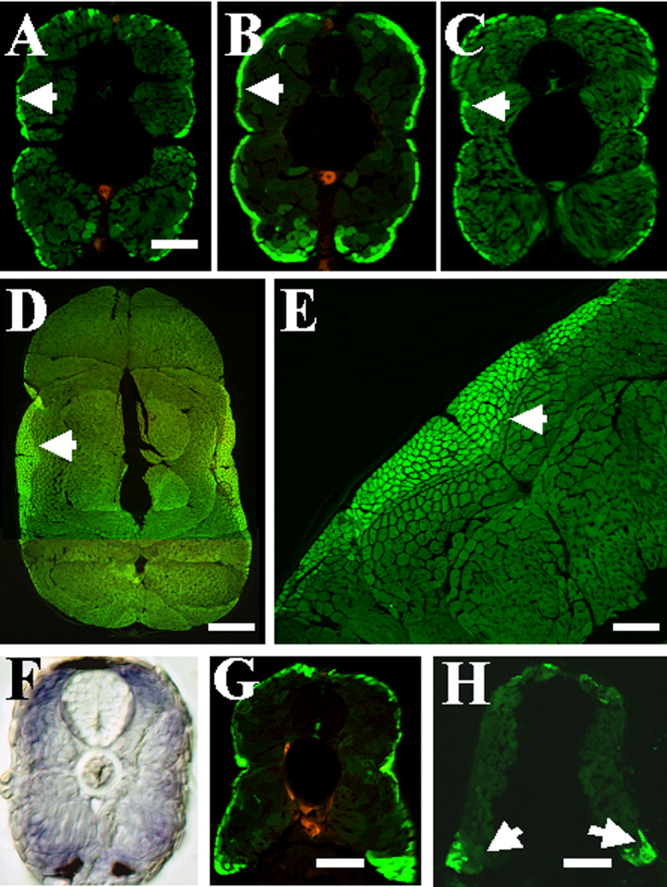
Fig. 5 Different levels of green fluorescent protein (GFP) expression in slow and fast muscles of smyd1-gfp transgenic fish. A-C: GFP expression directly photographed on embryonic sections at 4 days postfertilization (dpf) under a confocal microscope. Slow muscles express higher levels of GFP than fast muscles in three transgenic lines of smyd1-gfp transgenic fish. A, line-27; B, line-32; C, line-51. Arrows indicate slow muscles. D,E: Cross-section showing higher levels of GFP expression in slow muscles of line-32 at 2 months old. Slow muscles are indicated by arrows. F: Cross-section shows smyd1 mRNA expression in both slow and fast muscles at 4 dpf, although the staining appeared slightly stronger in superficial slow muscles. G,H: Cross-sections (dorsal on top) showing GFP expressing slow muscles in wild-type transgenic larvae (G) or yot mutant larvae (H) at 4 dpf. Slow muscles are clearly present at the dorsal and ventral myotome in yot mutant embryos. Scale bars = 150 μm in A-C, 500 μm in D, 200 μm in E, 120 μm in G,H.
Image
Figure Caption
Figure Data
Acknowledgments
This image is the copyrighted work of the attributed author or publisher, and
ZFIN has permission only to display this image to its users.
Additional permissions should be obtained from the applicable author or publisher of the image.
Full text @ Dev. Dyn.

