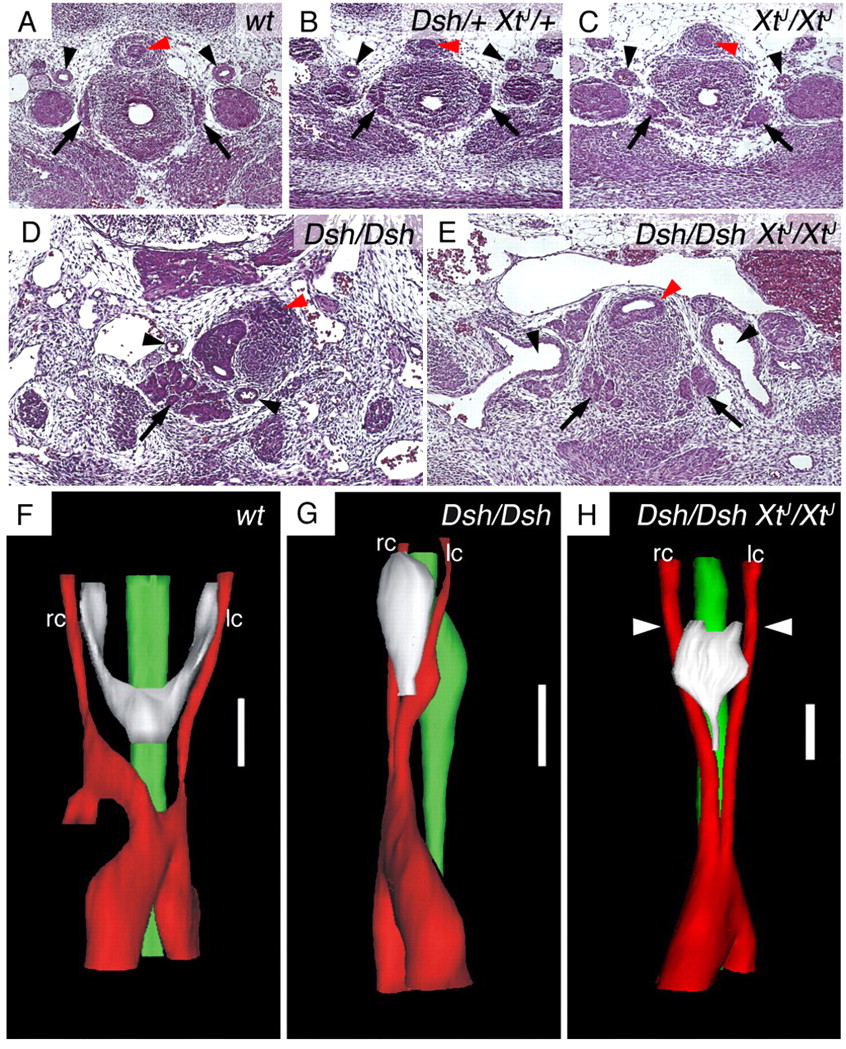
Fig. 5 In Dsh/Dsh XtJ/XtJ double mutants, symmetry of carotid arteries is restored and the thyroid primordium regains its midline position. HE staining of representative histological sections (A-E) and three-dimensional reconstruction (F-H) of E13.5 mouse embryos. (A-C) Both carotid arteries (arrowheads) and thyroid lobes (arrows) are normal in double heterozygous or XtJ/XtJ mouse embryos. (D,E) Whereas in Dsh/Dsh mice both carotid arteries and mis-shaped thyroid are positioned unilaterally (D), a bilateral set of carotid arteries (arrowheads) forms in double homozygous Dsh/Dsh XtJ/XtJ mice (E), and the thyroid develops into a bilateral set of lobes at its cranial end (arrows). The carotid arteries in E are larger than in wild type but their identity is unequivocally due to their arterial walls and origin from the aortic arch (as revealed by series of sections, data not shown). Red arrowheads indicate the oesophagus. A-E are at the same magnification; D,E show larger areas. (F-H) Three-dimensional reconstruction visualising how the carotid arteries and the thyroid have regained bilateral positions in Dsh/Dsh XtJ/XtJ mice. White arrowheads indicate the level of the section in E, showing the symmetrical cranial lobes of the thyroid. The thyroid is still abnormally shaped, probably owing to the close distance between the carotid arteries. lc, left carotid artery; rc, right carotid artery. Scale bars: 140 μm.
Image
Figure Caption
Acknowledgments
This image is the copyrighted work of the attributed author or publisher, and
ZFIN has permission only to display this image to its users.
Additional permissions should be obtained from the applicable author or publisher of the image.
Full text @ Development

