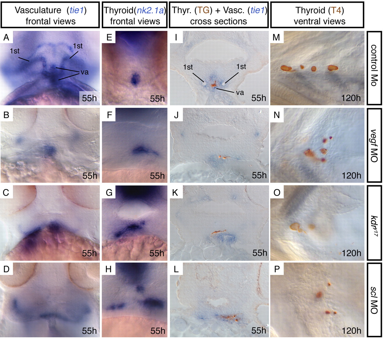
Fig. 2 Mutants with defects in ventral aorta development show correlating thyroid abnormalities. (A-H) Frontal views, showing vasculature (tie1 expression) or thyroid (nk2.1a expression). Control morpholino injected embryos are indistinguishable from wild-type embryos or kdry17 siblings. (I-L) Sections showing folliclular lumen (thyroglobulin immunostaining, brown) in relation to endothelial cells (tie1 expression, blue). (M-P) At 120 hpf, thyroid follicles (T4 immunostaining, brown) fail to align along the ventral aorta. In F-H,J-L the thyroid appears larger owing to the lateral expansion. However, in wild-type embryos the thyroid extends along the AP axis (compare with Fig. 1B). Concomitant with lateral expansion, its AP extent appears to be reduced, so that the size remains similar. This is also reflected by normal follicle numbers at 120 hpf (M-P). TG, thyroglobulin; Thyr, thyroid; Vasc, vasculature; va, ventral aorta; 1st, first pair of branchial arteries.
Image
Figure Caption
Figure Data
Acknowledgments
This image is the copyrighted work of the attributed author or publisher, and
ZFIN has permission only to display this image to its users.
Additional permissions should be obtained from the applicable author or publisher of the image.
Full text @ Development

