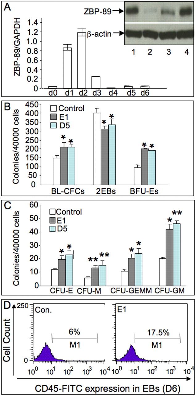
Fig. 6 Expression profile of ZBP-89 and effects of its overexpression on hematopoiesis in mouse EB cultures. (A) ZBP-89 expression profile in undifferentiated ESCs and differentiating EBs quantified with real-time PCR. Numbers indicate the day of differentiation. Results represent meanąs.d. of three independent experiments. Inset, western blot analysis showing induction of the ZBP-89 protein mainly in FLK1+ mesoderm precursors in day 3 EBs. Lane 1, positive control; lane 2, uninduced ESCs; lane 3, day 3 FLK1- mesodermal cells; lane 4, FLK1+ mesoderm precursors. Equivalent amounts of cell lysate were loaded per lane as reflected by the β-actin signal. (B) Histograms showing the number of blasts (BL-CFCs), secondary EBs (2°EBs) and primitive erythroid (BFU-Es) colonies generated from control, D5 and E1 clones (bars represent the mean number of coloniesąs.d. from two independent experiments). (C) Histograms (meanąs.d., n=3) showing the numbers of definitive erythroid (CFU-E), macrophage (CFU-M), granulocyte-macrophage-megakaryocyte (CFU-GEMM) and granulocyte-macrophage (CFU-GM) colonies. *P<0.01; **P<0.001 (paired t-test). (D) Flow cytometric analysis of a single-cell suspension from E1-derived EBs (see Materials and methods) stained with FITC-labeled rat antimouse CD45 monoclonal antibody. ZBP-89 overexpression significantly increased the number of CD45+ hematopoietic progenitors.
Image
Figure Caption
Acknowledgments
This image is the copyrighted work of the attributed author or publisher, and
ZFIN has permission only to display this image to its users.
Additional permissions should be obtained from the applicable author or publisher of the image.
Full text @ Development

