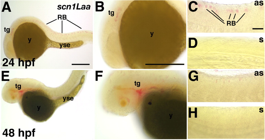
Fig. 2 Scn1Laa is expressed in sensory neurons of the peripheral nervous system and Rohon-Beard (RB) cells. A-D: At 24 hours postfertilization (hpf), scn1Laa mRNA is detected in the trigeminal ganglion and RB cells. A: A low-magnification view reveals expression in an anterior domain caudal to the eye and in RB cells throughout the spinal cord. B: At higher magnification, the anterior expression domain is recognized as the developing trigeminal ganglion migrating rostrally toward the eye. C: The posterior expression is found in RB cells of the spinal cord. D: The sense probe (s) does not reveal a signal under the same conditions used for the antisense probe (as). E-H: At 48 hpf, in situ hybridization signals are stronger in the trigeminal ganglion but weaker posteriorly in the spinal cord. E,F: At 48 hpf, the trigeminal ganglia have migrated to their characteristic position just caudal to the eye and continue to express scn1Laa. G: At 48 hpf, dorsal RB cells continue to express scn1Laa transcripts. H: The sense probe reveals no hybridization signal. as, antisense probe; n, notochord; RB, Rohon-Beard cell; s, sense probe; tg, trigeminal ganglion; yse, yolk sac extension. In this and subsequent figures, whole-mount photos are oriented with anterior to the left and dorsal up. Scale bars = 250 μm in A (applies to A,E), 100 μm in B (applies to B,F), 100 μm in C (applies to C,D,G,H).
Image
Figure Caption
Figure Data
Acknowledgments
This image is the copyrighted work of the attributed author or publisher, and
ZFIN has permission only to display this image to its users.
Additional permissions should be obtained from the applicable author or publisher of the image.
Full text @ Dev. Dyn.

