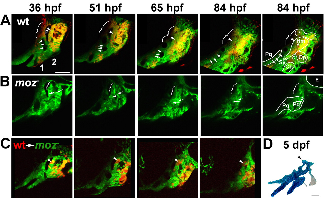
Fig. 6 Moz is required for the growth and chondrification of dorsal second-segment CNC. (A-C) Progressive frames, as indicated, from time-lapse confocal recordings of first (1) and second (2) pharyngeal segment development in wild-type fli1:GFP (A), moz-; fli1:GFP (B), and moz-; fli1:GFP/wild-type fli1:GFP mosaic (C) animals. Skeletal outlines are shown at 84 hpf. (A) A subset of CNC precursors are labelled with a red dye. In wild type (n=4), dorsal second-segment CNC (arrowheads) adjacent to p1 endoderm (P) form the anterior part of Hm cartilage. Dorsal first-segment CNC (brackets) form either no cartilage or neurocranial cartilage (*). Intermediate first-segment CNC (arrows) contribute to Pq cartilage. (B) In moz-; fli1:GFP animals (n=6), intermediate second-segment CNC contribute to Pq' cartilage (arrows). More dorsal CNC make no cartilage (brackets). In C, small amounts of wild-type CNC precursors (red) were transplanted into moz-; fli1:GFP animals. Wild-type second-segment CNC (arrowheads) adjacent to p1 grow and form a cartilage nodule in the normally cartilage-free dorsal zone of the moz- host. (D) The skeletal preparation of this same animal at 5 dpf. Scale bars: 50 Ám. See also Movies 1-3 in the supplementary material.
Image
Figure Caption
Acknowledgments
This image is the copyrighted work of the attributed author or publisher, and
ZFIN has permission only to display this image to its users.
Additional permissions should be obtained from the applicable author or publisher of the image.
Full text @ Development

