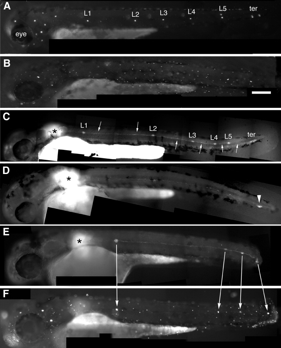
Fig. 4 Embryonic posterior lateral line labeled by 4-Di-2-ASP, a marker of differentiated hair cells (A, B, and F) or by uncaging the primordium (C-E). A: Wild type embryo with the normal pattern of five neuromasts along the horizontal myoseptum (L1-L5) and two terminal neuromasts (ter). B: Morphant embryo where all PLL neuromasts are missing. C: Uncaging the primordium at the position marked by the asterisk in a wild type embryo reveals the neuromasts (L1-5 and ter) and interneuromastic cells (arrows). D: Uncaging in a morphant reveals that the primordium has reached the tip of the tail (arrowhead) but no neuromasts have been deposited. E,F: In a partial morphant where four clusters have been deposited, including a terminal one (E), each cluster has differentiated hair cells on the next day (F).
Image
Figure Caption
Figure Data
Acknowledgments
This image is the copyrighted work of the attributed author or publisher, and
ZFIN has permission only to display this image to its users.
Additional permissions should be obtained from the applicable author or publisher of the image.
Full text @ Dev. Dyn.

