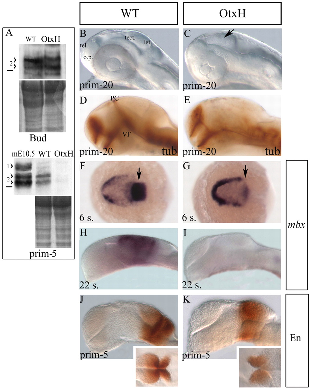
Fig. 1 Absence of mesencephalon in OtxH embryos. (A) Western blot analysis on wild-type or OtxH zebrafish embryo extracts from end of gastrulation stage (upper panel: bud stage) or late somitogenesis stage (lower panel: prim-5). Proteins staining is shown underneath each to control protein levels between wild-type and OtxH embryos. 1>, mouse Otx1protein; 2>, doublet of mouse or zebrafish Otx2 proteins. The horizontal black line represents the 36.4 kDa marker. In OtxH embryos, Otx2 expression is not visibly affected at the end of epiboly but is lost by the end of somitogenesis. (B-E) Lateral views, anterior towards the left, of prim-20 (33 hpf) live brains (B,C) or fixed after acetylated tubulin staining (D,E) from wild-type (B,D) or OtxH (C,E) embryos. The isthmic constriction (arrow in C), the ventral flexure (VF) and the posterior commissure (PC, normally forming at the boundary between forebrain and midbrain) are all absent. tel, telencephalon; o.p., olfactory placode; tect., tectum; ist, isthmus. Dorsal (F,G) and lateral (H-K) views, anterior towards the left, of wild-type (F,H,J) or OtxH (G,I,K) brains. Expression of mbx is almost undetectable in the CNS at the six-somite stage in OtxH embryos (arrow in G) compared with wild type (arrow in F) and is lost by the 22-somite stage (I). Expression of Engrailed is detectable in both wild type (J) and OtxH (K) at prim-5 stage. Insets are dorsal views of the isthmic area of the brains shown in J and K.
Image
Figure Caption
Figure Data
Acknowledgments
This image is the copyrighted work of the attributed author or publisher, and
ZFIN has permission only to display this image to its users.
Additional permissions should be obtained from the applicable author or publisher of the image.
Full text @ Development

