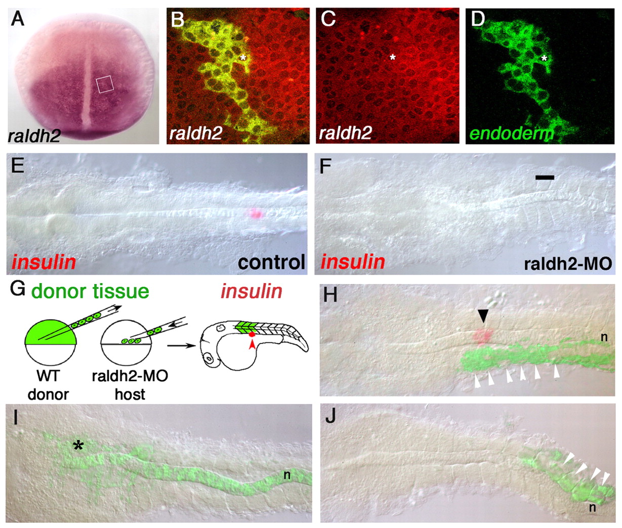
Fig. 1 The RA required for pancreas development is produced in anterior paraxial mesoderm. (A) raldh2 expression in dorsal view (anterior to top) at 10 hpf. (B-D) raldh2 expression in endoderm cells; an example is indicated with an asterisk. Confocal images of raldh2 expression (red) at 10 hpf, SOX32-positive donor-derived endoderm is labeled with fluorescein dextran (green). Approximate area of image is boxed in A. (E,F) insulin expression (red) at 24 hpf. (E) Normal insulin expression. (F) No insulin expression appears in raldh2 morpholino injected embryos (bar indicates normal position of pancreas; 100%; n=38). (G) Schematic of cell-transplantation approach. Fluorescein dextran-labeled donor cells from sphere stage (4 hpf) embryos are distributed along the blastoderm margin of raldh2-MO injected hosts. Donor cells contribute to mesoderm (indicated here in anterior somites). (H-J) Transplanted, raldh2-MO injected embryos probed for insulin expression at 24 hpf (red). Photographs are composites of bright field and fluorescent images to detect donor tissue (green). (H) An embryo in which donor tissue contributed to anterior somites (white arrowheads), rescuing insulin expression (arrowhead). (I,J) Embryos in which donor tissue contributed to notochord (n) plus overlying neural cells (*) (I) and posterior somites (white arrowheads) (J); no insulin expression is detectable.
Image
Figure Caption
Figure Data
Acknowledgments
This image is the copyrighted work of the attributed author or publisher, and
ZFIN has permission only to display this image to its users.
Additional permissions should be obtained from the applicable author or publisher of the image.
Full text @ Development

