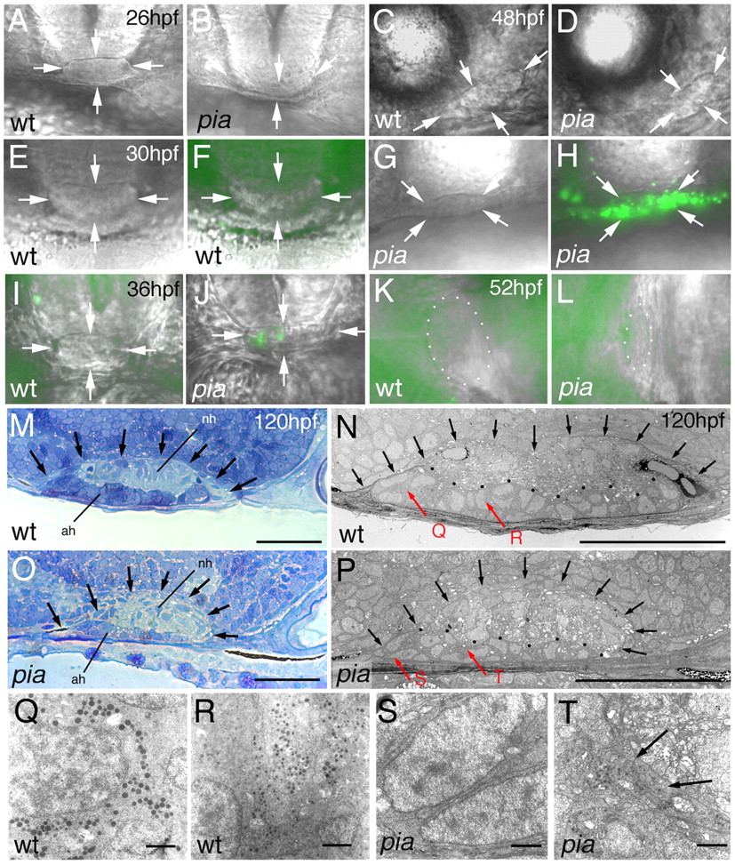
Fig. 4 pia mutants display indistinct early pituitary morphology, followed by a transient phase of adenohypophyseal cell death and the formation of a smaller, but distinct, pituitary gland with cells of rather primitive ultrastructure. (A-L) Nomarski images of live pia mutant embryos (pia) and their corresponding wild-type siblings (wt). (F,H,I-L) Images are superimposed by Acridine Orange staining for apoptosis. (A,B,E-J) Frontal views, dorsal upwards; (C,D) lateral views, anterior towards the left, dorsal upwards; (K,L) ventral views, anterior towards the right. Ages of embryos are indicated in upper right-hand corners of wild types. Genotypes were determined via PCR after photography. Arrowheads in A-J indicate borders of the pituitary gland; in K,L, pituitary borders are outlined by dots. (M-T) Pituitary ultrastructure at 120 hpf; longitudinal sections, anterior towards the left, dorsal towards the top. (M,O) Toluidine Blue staining; (N,P-T) electron micrographs. (M-P) The border of the pituitary is indicated by arrows; (N,P) the border between adenohypophysis (ah) and neurohypohysis (nh) is outlined by black dots. (Q-T) Higher magnifications of regions indicated by red arrows in N,P. Vesicles (as in T, indicated by arrows) were seen in three out of ~20 adenohypophyseal cells present in the section of the pia pituitary (P). They could contain matrix proteins and hormone-binding proteins, which can be made even in the absence of hormone production (compare with Norris, 1997). Scale bars: 30 μm in M-P; 1 μm in Q-T.
Image
Figure Caption
Acknowledgments
This image is the copyrighted work of the attributed author or publisher, and
ZFIN has permission only to display this image to its users.
Additional permissions should be obtained from the applicable author or publisher of the image.
Full text @ Development

