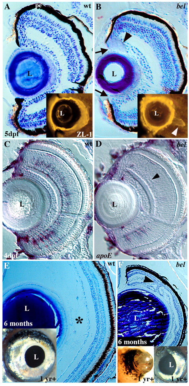
Fig. 7 Larval and adult eye defects in bel mutants. (A, B) Toluidine Blue stained sections from 5 dpf wild type (A) and bel mutants (B). In bel mutants there is a gap between the PE and lens (arrows), and an abnormal acellular mass is often present (arrowhead). (Inset) In a different embryo, an acellular aggregate was labeled with the ZL-1 antibody, indicating it contains lens proteins (arrowhead). (C,D) At 4 dpf, bel mutants (D) lack most amacrine cells, as determined by apoE in situ labeling (arrowhead). A few cells remain in the ventral retina. (E, F) Eyes of surviving bel adults (F) are much smaller than those of wild type (E) due to the lack of a posterior compartment (asterisk). Retinal layers are folded back on one another (arrowhead). Insets show lateral views of whole eyes. (F, left inset) In some adults, vascularized retinal tissue extends out of the eye. L, lens; pe, pigmented epithelium.
Image
Figure Caption
Acknowledgments
This image is the copyrighted work of the attributed author or publisher, and
ZFIN has permission only to display this image to its users.
Additional permissions should be obtained from the applicable author or publisher of the image.
Full text @ Development

