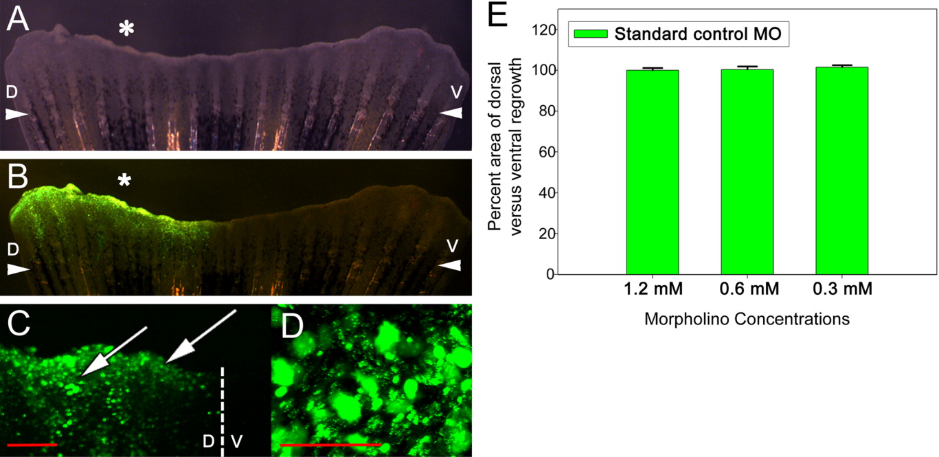
Fig. 2 Injection and in vivo electroporation of standard control morpholino. A: Bright-field image of injected and electroporated fin at 24 hpa. B: Fluorescence image of the same injected and electroporated fin at 24 hpa. C: Confocal Z-stack of a region of an electroporated fin at 24 hpa showing fluorescence in both the underlying mesenchyme and overlying wound epidermis (arrows). The dotted line denotes the boundary between the dorsal (D) and ventral (V) sides of the fin. D: Confocal Z-stack of a magnified region, proximal to the epidermis and distal to the end of a bony ray, of the dorsal side of the fin shown in C. E: Graph comparing areas of outgrowth of dorsal (experimental) and ventral (control) sides at 72 hpa. At least five fins were examined for each concentration of morpholino. *, injected dorsal side of the fin; arrowheads, plane of amputation; D, dorsal side; V, ventral side. Scale bar (red) in C = 200 μm and in D = 50 μm.
Image
Figure Caption
Acknowledgments
This image is the copyrighted work of the attributed author or publisher, and
ZFIN has permission only to display this image to its users.
Additional permissions should be obtained from the applicable author or publisher of the image.
Full text @ Dev. Dyn.

