Image
Figure Caption
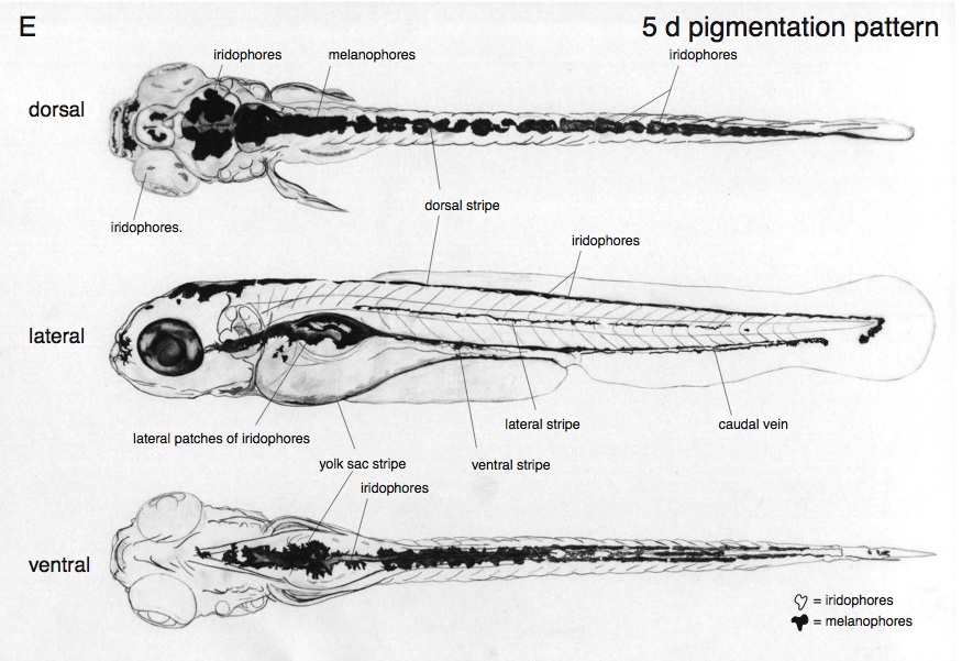
Fig. 2E Drawings of embryos at 24 hours (A), 48 hours (B,D), and 5 days (C,E) of development. For clarity, the melanophore pigmentation pattern is omitted from B and C. It is depicted in D and E. Most of the structures that can be seen in the living embryo with a compound microscope are marked.
Acknowledgments
This image is the copyrighted work of the attributed author or publisher, and
ZFIN has permission only to display this image to its users.
Additional permissions should be obtained from the applicable author or publisher of the image.
Full text @ Development

