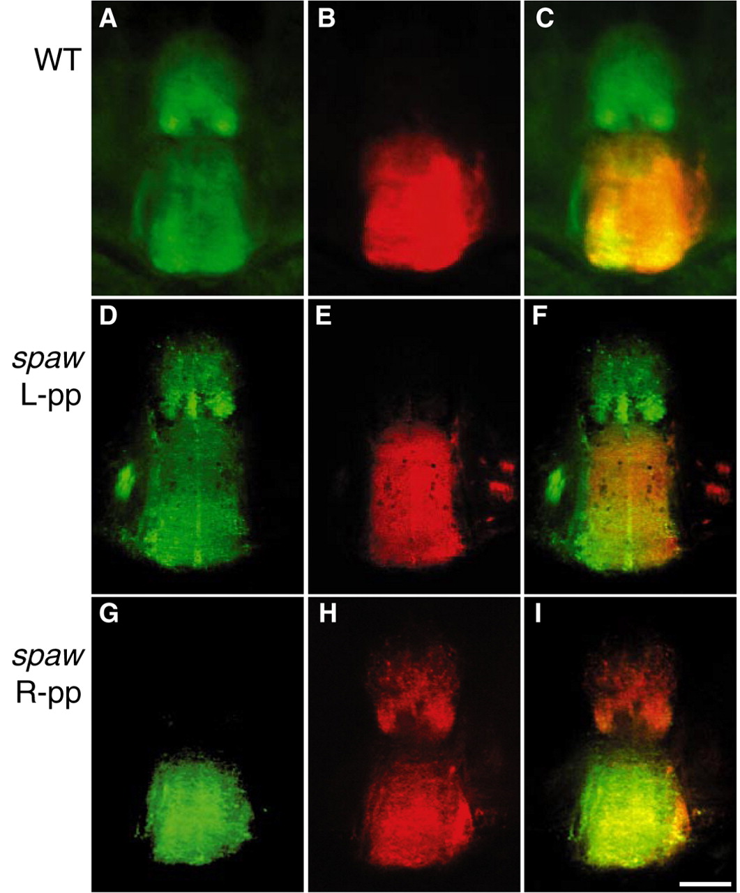
Fig. 5 Directional asymmetry influences IPN projection pattern. Habenular projections onto the IPN in sections (150 Ám) through adult midbrain of (A-C) WT and (D-I) spaw MO-injected fish, which as larvae had a left (D-F) or right (G-I) positioned parapineal. (A-F) Axons originating from left habenula innervate dorsal and ventral IPN domains, while those from the right habenula only project ventrally. (G-I) In fish with reversed epithalamic laterality, L-R origin of IPN projections is also reversed, such that axons from left habenula now innervate only the ventral IPN. A-C are higher magnification Leica MZFLIII images of the same brain as in Fig. 4. D,E and G,H were imaged using a Leica SP2 confocal microscope and merged to produce the composite images in F,I. Dorsal is at the top in all images. Scale bar: 60 Ám.
Image
Figure Caption
Acknowledgments
This image is the copyrighted work of the attributed author or publisher, and
ZFIN has permission only to display this image to its users.
Additional permissions should be obtained from the applicable author or publisher of the image.
Full text @ Development

