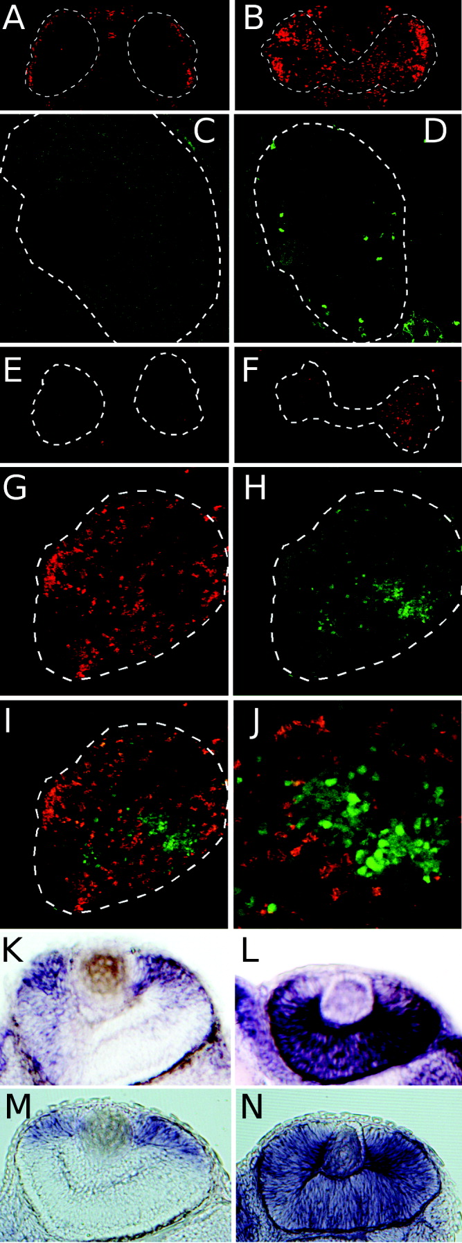
Fig. 4 >Loss of hdac1 activity results in failure of retinal cells to exit the cell cycle. A,B: A 2-hr bromodeoxyuridine (BrdU) incorporation in 72 hr postfertilization (hpf) wild-type (A) or hdac1 mutant (B) embryos. C,D: Anti-H3 immunofluorescence on cryosections of 72 hpf wild-type (C) or hdac1 mutant (D) eyes. E,F: Terminal deoxynucleotidyl transferase-mediated deoxyuridinetriphosphate nick end-labeling (TUNEL) staining of apoptotic cells in 72 hpf wild-type (E) and hdac1 mutant (F) embryos. G-J: Wild-type green fluorescent protein (GFP) -expressing cells (green, H,I) were transplanted into hdac1 mutant embryos. BrdU incorporation is shown (red, G,I). J: Higher magnification of the image shown in I. A-J shows immunostaining on cryosections, anterior up. K-N: Whole-mount in situ hybridizations of cyclin D1 RNA (K,L), and cyclin E2 RNA (M,N); 48 hpf wild-type (K,M) and hdac1 mutant (L,N) embryos (anterior right, dorsal up)
Image
Figure Caption
Figure Data
Acknowledgments
This image is the copyrighted work of the attributed author or publisher, and
ZFIN has permission only to display this image to its users.
Additional permissions should be obtained from the applicable author or publisher of the image.
Full text @ Dev. Dyn.

