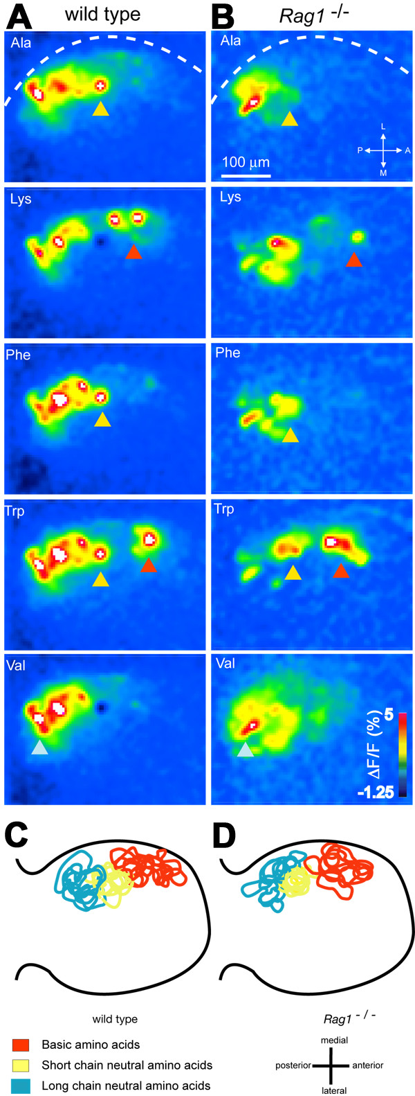
Fig. 6 Chemotopic organization of glomerular activity patterns in wild type and Rag1 mutant fish. (A) Glomerular activity patterns evoked by different amino acid stimuli (10-5 M) in the ventro-lateral OB of a wild type fish. Changes in fluorescence intensity report activity of OSN axon terminals and are colour coded. Dashed line depicts lateral edge of the OB. Arrowheads indicate conserved clusters of glomeruli with defined response properties. (B) Glomerular activity patterns evoked by the same stimuli in a Rag1 mutant fish. The same general areas of the bulb show a response. The difference in the intensity seen here between wild type and mutants is not statistically significant. (C) Overlay of positions of identifiable glomerular regions in wild type of the line from which Rag1 mutants were derived (n = 7). Regions were outlined manually in activity maps in each fish. Outlines from different individuals were centered on the central cluster (yellow). (D) Overlay of cluster positions determined in 10 Rag1 mutant fish.
Image
Figure Caption
Acknowledgments
This image is the copyrighted work of the attributed author or publisher, and
ZFIN has permission only to display this image to its users.
Additional permissions should be obtained from the applicable author or publisher of the image.
Full text @ BMC Neurosci.

