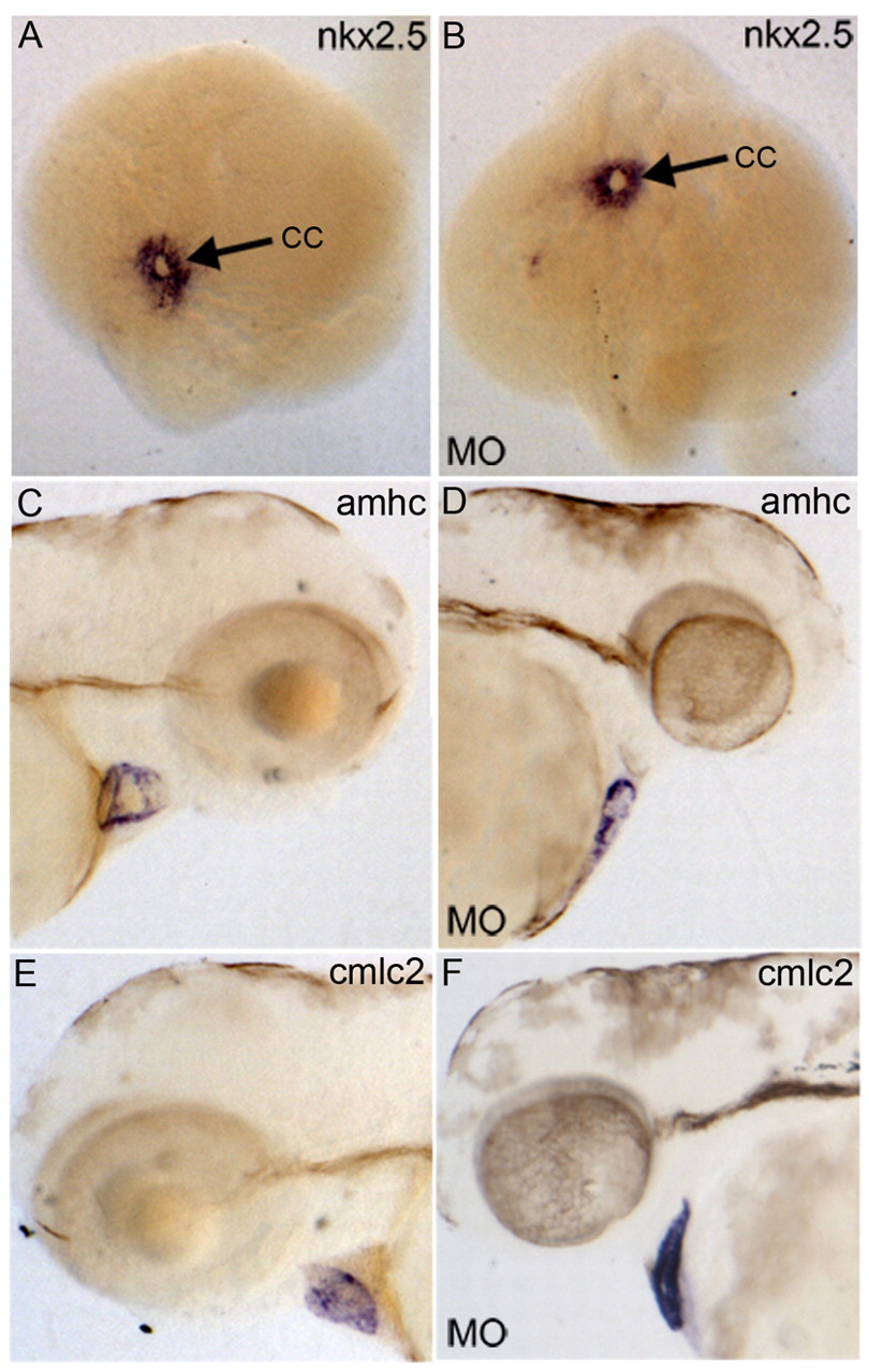
Fig. 3 Heart tube formation is normal, but morphogenesis fails in Gata4 deficient embryos. Control uninjected embryos (A,C,E) are compared with embryos derived from eggs injected with MO (B,D,F). Embryos were processed for in situ hybridization to detect transcripts for Nkx2.5 (A,B), Amhc (C,D) or Cmlc2 (E,F). The morphant embryos show normal formation of the cardiac cone (cc) at the 20-somite stage (B). Likewise, the cardiomyocyte markers Amhc and Cmlc2 are expressed normally, shown here at 3 dpf. (A,B) Dorsal views with anterior to the top; (C,D) lateral views with anterior to the right and dorsal to the top; (E,F) lateral views with anterior to the left and dorsal to the top. Embryos are shown in both orientations to show the lack of looping in the linear heart tube of the morphant embryos.
Image
Figure Caption
Figure Data
Acknowledgments
This image is the copyrighted work of the attributed author or publisher, and
ZFIN has permission only to display this image to its users.
Additional permissions should be obtained from the applicable author or publisher of the image.
Full text @ Development

