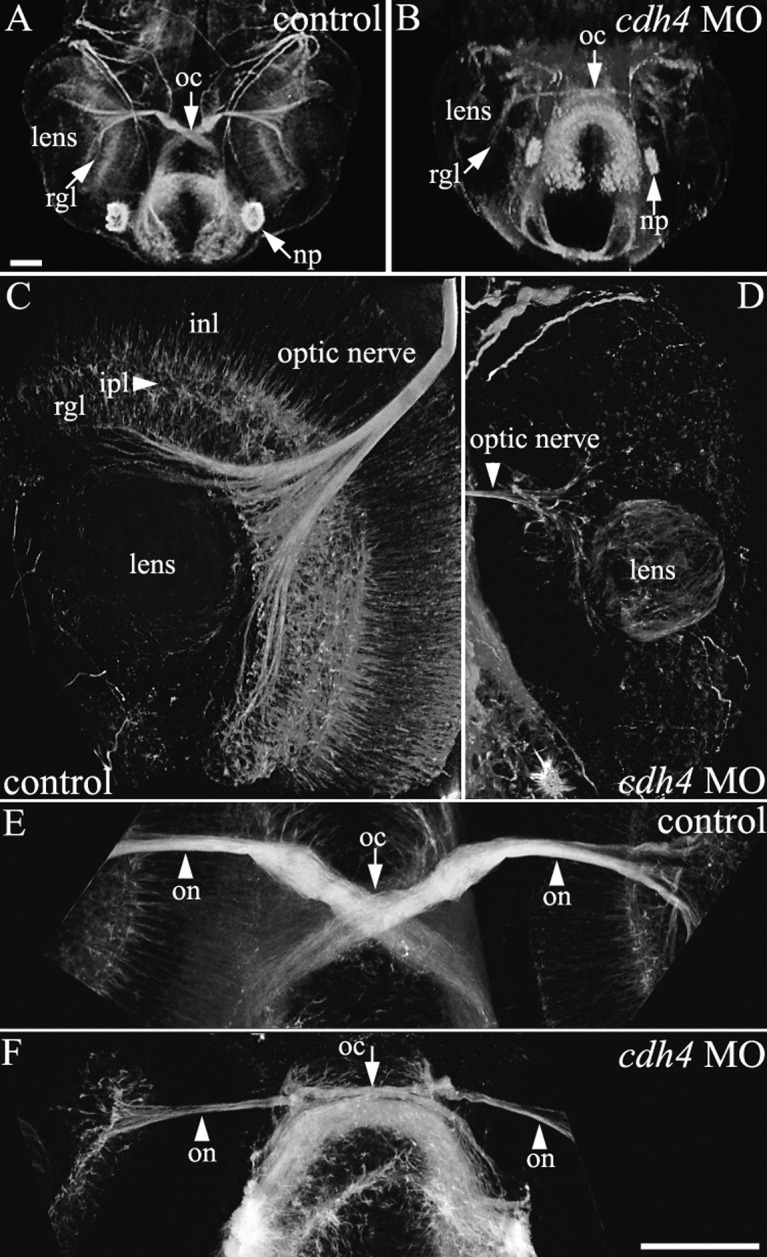
Fig. 4 cdh4 knockdown results in sparse retinal ganglion cell axon outgrowth at 2 days postfertilization (dpf). A-F: Zebrafish embryos were injected with either standard control morpholino oligonucleotide (MO, A,C,E) or cdh4 MO (B,D,F). Embryos were fixed at 2 dpf, and whole-mount preparations with axon tracts immunostained using acetylated tubulin antibodies were visualized using two-photon imaging. Ventral view projections are shown of the planes containing the eyes and optic nerves. A,B: Low-magnification views are shown for orientation. A: In control embryos, axons exit the retinal ganglion cell layer to form the optic nerve, which then bends to travel more dorsally and rostrally, crosses the midline at the optic chiasm, then projects straight toward the contralateral optic tectum. B: In severe cdh4 knockdowns, both the retinal ganglion cell layer and optic nerve are sparse and the bend in the optic nerve as it travels toward the optic chiasm is absent, but the sparse optic nerve does project normally across the midline toward the contralateral optic tectum. C,D: In high-resolution views of the control eye, extensive neural processes of all the major retinal layers are evident (C), whereas in the cdh4 knockdown eye, neuronal processes are largely absent and only a sparse disorganized retinal ganglion cell layer is visible (D). Projection images of two-photon image volumes are shown. E,F: High-resolution views of the optic chiasm show the thick well fasciculated optic nerve of the control (E) and the thin loosely fasciculated optic nerve of the cdh4 knockdown (F). Scale bars = 50 μm.
Image
Figure Caption
Figure Data
Acknowledgments
This image is the copyrighted work of the attributed author or publisher, and
ZFIN has permission only to display this image to its users.
Additional permissions should be obtained from the applicable author or publisher of the image.
Full text @ Dev. Dyn.

