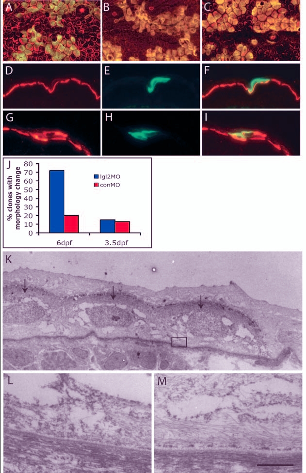
Fig. 6 Clones derived from antisense lgl2 morpholino (lgl2MO)-injected embryos recapitulate the pen phenotype. (A-I) Confocal and immunohistological analysis of recipient embryos with epidermal clones labelled with GFP (green) and keratin (red). In overlays (A-C,F,I), clones appear yellow. At 6 dpf, clones derived from control morpholino (conMO)-injected embryos (A) comprise polygonal and flattened cells, similar to wild-type epidermal cells, while those derived from lgl2MO-injected embryos (B) showed changes in cellular morphology, becoming spindle or round shaped, similar to pen mutant. By contrast, on 3.5 dpf, clones containing lgl2MO (C) did not show any changes in cellular morphology. Immunohistological analysis of clones (6dpf) with conMO (D-F) reveals basal localisation of keratin, whereas clones containing lgl2MO exhibit mislocalisation of keratin (G-I). (J) Quantification of clones containing conMO or lgl2MO and exhibiting pen-like phenotype. Small proportions of clones carrying conMO comprise cells that deviate from usual morphology or show spindle shapes. There is a vast increase in the proportion of clones exhibiting pen-like phenotype at 6 dpf but not at 3.5 dpf (when, instead, they carry lgl2MO). (K-M) Electron microscopic analysis of hemidesmosome formation. Clone marked with GFP is detected with electron-dense DAB (arrows in K). Hemidesmosomes are absent in the clone (L represents boxed region in K) in contrast to the control recipient epidermis (M). Scale bar: 7 Ám in K; 543 nm in L,M.
Image
Figure Caption
Acknowledgments
This image is the copyrighted work of the attributed author or publisher, and
ZFIN has permission only to display this image to its users.
Additional permissions should be obtained from the applicable author or publisher of the image.
Full text @ Development

